. 2016 Aug;74(6):ftw058.
doi: 10.1093/femspd/ftw058. Epub 2016 Jun 14.

The Rickettsia type IV secretion system: unrealized complexity mired by gene family expansion

Affiliations

The Rickettsia type IV secretion system: unrealized complexity mired by gene family expansion

Joseph J Gillespie et al. Pathog Dis. 2016 Aug.

Abstract

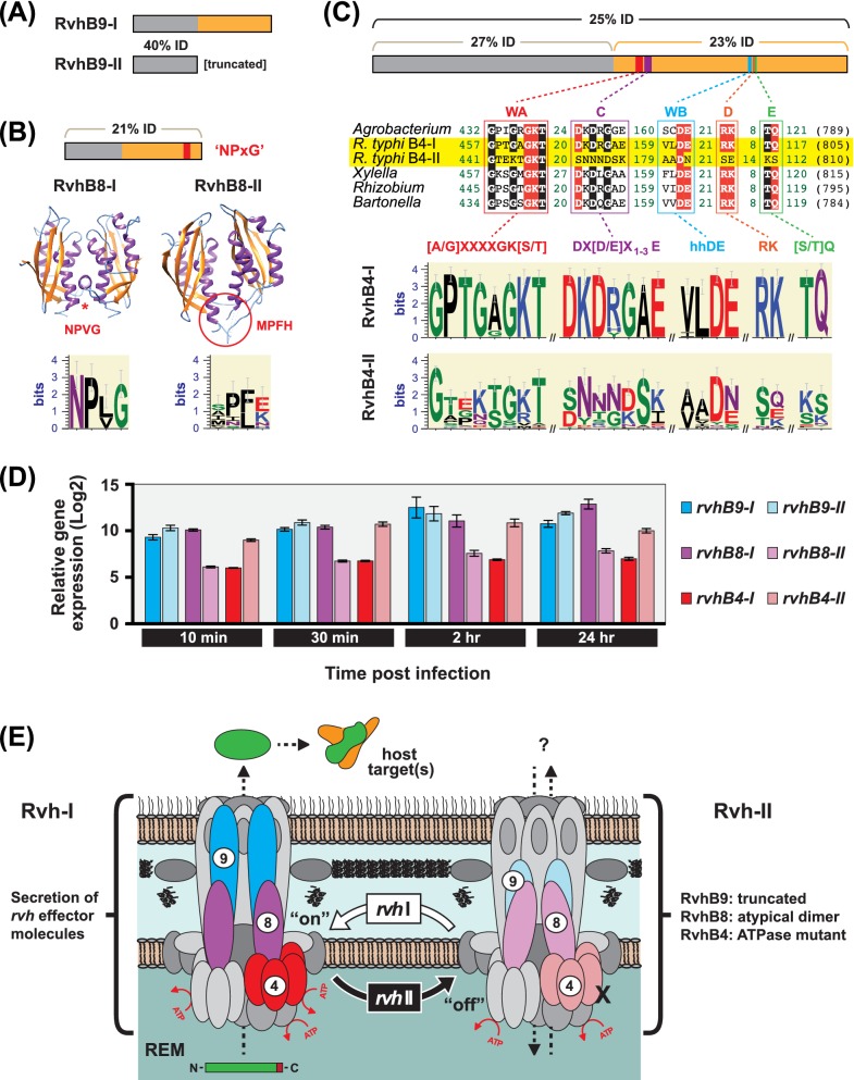
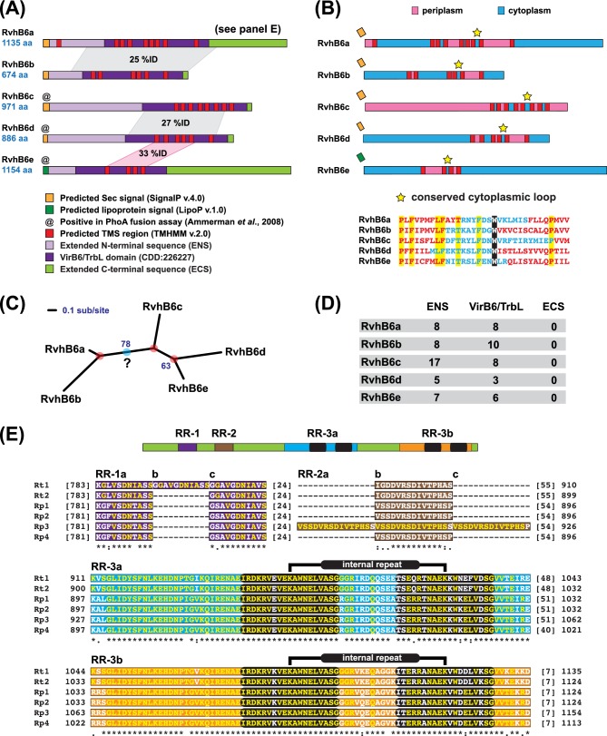
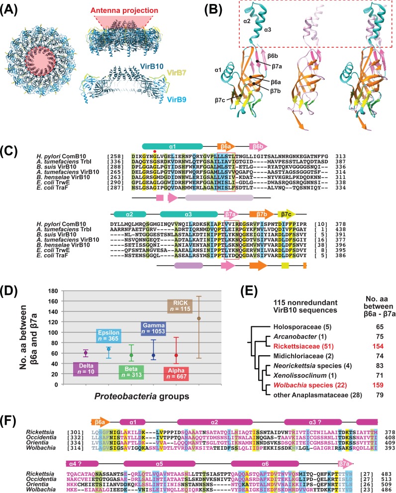
Many prokaryotes utilize type IV secretion systems (T4SSs) to translocate substrates (e.g. nucleoprotein, DNA, protein) across the cell envelope, and/or to elaborate surface structures (i.e. pili or adhesins). Among eight distinct T4SS classes, P-T4SSs are typified by the Agrobacterium tumefaciens vir T4SS, which is comprised of 12 scaffold components (VirB1-VirB11, VirD4). While most P-T4SSs include all 12 Vir proteins, some differ from the vir archetype by either containing additional scaffold components not analogous to Vir proteins or lacking one or more of the Vir proteins. In a special case, the Rickettsiales vir homolog (rvh) P-T4SS comprises unprecedented gene family expansion. rvh contains three families of gene duplications (rvhB9, rvhB8, rvhB4): RvhB9,8,4-I are conserved relative to equivalents in other P-T4SSs, while RvhB9,8,4-II have evolved atypical features that deviate substantially from other homologs. Furthermore, rvh contains five VirB6-like genes (rvhB6a-e), which are tandemly arrayed and contain large N- and C-terminal extensions. Our work herein focuses on the complexity underpinned by rvh gene family expansion. Furthermore, we describe an RvhB10 insertion, which occurs in a region that forms the T4SS pore. The significance of these curious properties to rvh structure and function is evaluated, shedding light on a highly complex T4SS.

Keywords: Rickettsia; Rickettsiales vir homolog; gene duplication; obligate intracellular bacteria; pathogenesis; type IV secretion system.


Figures

Figure 1.
Figure 1.
The rvh T4SS deviates from the P-T4SS archetype. A general model of P-T4SSs is shown, with a description for the functions of all 12 components (VirB1-VirB11, VirD4). A comparison with the rvh T4SS is provided, with deviations colored red. Inset (gray box) depicts the distribution of the rvh gene clusters in the R. typhi str. Wilmington genome (NCBI RefSeq NC_006142). Individual genes are uniform and not drawn to scale.
Figure 2.
Figure 2.
The nature of rvh gene duplication. (A–C) Specific characteristics of rvh paralogs. For each protein schema, NTDs and CTDs are colored gray and orange, respectively. Sequence logos were generated using WEBLOGO v.3.3 (Crooks et al.2004). (A) Rickettsia RvhB9 paralogs differ in domain architecture. Schema shows %ID between R. typhi RvhB9-I and RvhB9-II NTDs; RvhB9-II lacks the entire CTD (Gillespie et al.2009). (B) RvhB8 paralogs are structurally divergent. Schema shows %ID between R. typhi RvhB8-I and RvhB8-II, with approximation of the NPXG dimerization motif shown in red. Structural model (RvhB8-I) and crystal structure (RvhB8-II, PDBID: 4O3V) illustrate the divergent dimers formed by R. typhi RvhB8 paralogs, with emphasis on the NPXG motifs (red). Below each structure, sequence logos depict the composition of the NPXG motif across non-redundant RvhB8-I and RvhB8-II proteins from other species of Rickettsiales (Gillespie et al.2015b). (C) RvhB4 paralogs have divergent NTPase active sites. Schema shows %ID between R. typhi RvhB4-I and RvhB4-II. Five motifs that form the NTPase active site within the CTD are illustrated below across RvhB4-I and RvhB4-II (highlighted yellow) and VirB4 from four other proteobacterial species. Black, conserved residues; red, critical residues. Below the alignment, sequence logos depict the composition of the five conserved motifs across non-redundant Rickettsiales RvhB4-I and RvhB4-II proteins. For motifs above sequence logos: X, any residue; h, any hydrophobic residue. All alignments generated using MUSCLE (Edgar 2004) with default parameters. (D) Expression of R. typhi rvhB9, rvhB8 and rvhB4 genes during early host cell infection. RNA was extracted from HeLa cells infected with R. typhi and gene expression of RT0277 (encoding RvhB9-I, NCBI accession no. AAU03757), RT0281 (RvhB9-II, AAU03761), RT0280 (RvhB8-I, AAU03760), RT0278 (RvhB8-II, AAU03758), RT0033 (RvhB4-I, AAU03521) and RT0771 (RvhB4-II, AAU04227) was measured by reverse transcription quantitative PCR (RT-qPCR). Gene expression was normalized to R. typhi reference genes adr1 and sca5 (2ΔCT). Infections were repeated in triplicate with technical duplicate readings for RT-qPCR. Mean ± SEM is plotted. (E) Model for RvhB9,8,4 paralogs within the rvh T4SS. At left, Rvh-I depicts RvhB9,8,4-I paralogs as components of a secretion machine, which translocates substrates (rvh effector molecules). At right, Rvh-II depicts RvhB9,8,4-II paralogs as components of a transporter with unknown function (import or export of substrates). A working hypothesis for rvh autoregulation, wherein RvhB9,8,4 paralogs cycle on and off of a conserved rvh scaffold (RvhB2, RvhB3, RvhB6, RvhB7, RvhB10, RvhB11, RvhD4) to regulate secretion, is shown at center. The proliferated RvhB6 proteins are not illustrated in this model (see Fig. 3).
Figure 3.
Figure 3.
Characteristics of Rickettsia RvhB6 proteins. (A) Architectures of R. typhi RvhB6 proteins (RT0028-RT0032, NCBI accession nos. AAU03516–AAU03520). Gray shading between proteins indicates the significant alignments yielded by bidirectional BLASTP analysis between all five RvhB6 proteins. Pink shading depicts the best significant alignment to RvhB6e. (B) Putative membrane localizations for R. typhi RvhB6 proteins based on prediction of TMS regions using TMHMM v. 2.0 (Krogh et al.2001). Sequences of the conserved cytoplasmic loop are shown below: black, Trp residue shown to be required for polar localization of VirB6 in A. tumefaciens (Judd, Kumar and Das 2005); yellow, invariant residues across all RvhB6 proteins. (C) Phylogeny estimation of the conserved cytoplasmic loop region of R. typhi RvhB6 proteins. Maximum likelihood-based phylogeny was estimated with RAxML (Stamatakis 2014) using the WAG amino acid substitution model and implementing a gamma model of rate heterogeneity and estimation of the proportion of invariable sites. Branch support was assessed via 1000 bootstrap pseudoreplications. Red circles depict predicted duplication events, with the blue circle illustrating a hypothetical RvhB6 progenitor. (D) Number of Cys residues per domain for each of the R. typhi RvhB6 proteins. (E) RvhB6a homologs have a variable ECS. Schema (top) depicts the RvhB6a ECS for species of Typhus Group rickettsiae, with the positions of three RRs illustrated. Multiple sequence alignment (bottom) illustrates the structure of the RRs, with lowercase letters demarcating the specific repeat units. Yellow residues are identical across repeat units within the same sequence. Black ellipses within RR-3 illustrate internal repeats within each larger repeat. Taxon abbreviations and NCBI accession nos. are as follows: Rt1: R. typhi str. Wilmington (AAU03520), R. typhi str. TH1527 (AFE53897); Rt2: R. typhi str. B9991CWPP (AFE54735); Rp1: R. prowazekii str. BuV67-CWPP (AFE50617); Rp2: R. prowazekii str. Madrid E (NP_220496), R. prowazekii str. Chernikova (AFE48928), R. prowazekii str. Katsinyian (AFE49773), R. prowazekii str. Dachau (AFE51458), R. prowazekii str. NMRC Madrid E (AGJ01341), R. prowazekii str. Breinl (AGJ02751); Rp3: R. prowazekii str. GvV257 (AFE52553), R. prowazekii str. RpGvF24 (AFE53124); Rp4: R. prowazekii str. Rp22 (ADE29612), R. prowazekii str. Cairo 3 (EOB09562).
Figure 4.
Figure 4.
Rickettsia RvhB10 proteins harbor a large insertion. (A) Bird's-eye (left) and side (right) views of the tetradecameric P-T4SS CC encoded by plasmid pKM101 of E. coli (PDBID: 3JQO), adapted from Chandran et al. (2009). For the bottom-side view, seven heterodimers are not shown to provide a cross-sectional view. Colors for the CC subunits (VirB7, VirB9 and VirB10) are similar to the model in Fig. 1. The proximal location of the AP is highlighted red. (B) Two VirB10 family proteins are structurally divergent. Structures of H. pylori ComB10 (PDBID: 2BHV) (Terradot et al.2005) (left), E. coli TraF (PDBID: 3JQO) (Chandran et al.2009) (center) and both structures superimposed (right). Images generated with the UCSF Chimera package (Pettersen et al.2004). Dashed red box encloses the APs. (C) Multiple sequence alignment of diverse proteins of the VirB10 family. Alignment encompasses the region of the ComB10 labeled in panel B (helix α1 to strand β7c). The secondary structures of ComB10 and TraF are shown above and below the alignment, respectively, with helices and strands colored according to the structures in panel B. Red ball denotes the conserved Gly critical for gating the OM pore (Banta et al.2011). Invariant residues (yellow) and residues with conservation between groups of strongly similar (blue) or weakly similar (green) properties are highlighted. Alignment generated using MUSCLE (Edgar 2004) with default parameters. Full taxon names and NCBI accession nos. as follows: H. pylori ComB (NP_206842-NP_206843); A. tumefaciens TrbI (AAC82638); Brucella suis VirB10 (NP_699267); A. tumefaciens VirB10 (AAK90938); B. henselae VirB10 (CAF28107); E. coli TrwE (YP_009077459); E. coli TraF (YP_009074496). (D) Average lengths between strands β6a and β7a of VirB10 family proteins for five classes of Proteobacteria (n = 2408) and Rickettsiales (n = 115). All 2513 proteins were aligned with MUSCLE (default parameters) with the regions spanning strands β6a and β7a (boxed in panel C) extracted for computation. Bars denote ranges of lengths per group. (E) Large RvhB10 insertions are unique to two groups of Rickettsiales. Only species in the genera Rickettsia, Orientia and Occidentia (Rickettsiaceae) and all Wolbachia species were found to contain large insertions within the VirB10 AP. NOTE: all Holosporaceae species contain non-RvhB10 sequences (e.g. F-T4SS proteins). (F) RvhB10 insertions of Rickettsiaceae and Wolbachia species are predicted to be rich in alpha helices. Alignment generated using MUSCLE (default parameters), with a consensus secondary structure prediction shown above, as generated using JPred4 (Drozdetskiy et al.2015). JPred4 predictions for individual sequences are reflected by coloring (maroon, α-helices; gray, β-strands). Helices α3 and α4 are denoted with ?s, as alternative algorithms did not robustly support helices within these regions (data not shown). Residue highlighting as described in panel C. Full taxon names and NCBI accession nos. as follows: R. typhi str. Wilmington (YP_067244); Occidentia massiliensis str. Os18 (WP_019230977); Orientia tsutsugamushi str. Boryong (YP_001248092); Wolbachia endosymbiont of Drosophila melanogaster (NP_965840).

Similar articles

Cited by

References

    1. Al-Khedery B, Lundgren AM, Stuen S et al. . Structure of the type IV secretion system in different strains of Anaplasma phagocytophilum. BMC Genomics 2012;13:678. - PMC - PubMed
    1. Alvarez-Martinez CE, Christie PJ. Biological diversity of prokaryotic type IV secretion systems. Microbiol Mol Biol R 2009;73:775–808 - PMC - PubMed
    1. Ammerman NC, Rahman MS, Azad AF. Characterization of Sec-translocon-dependent extracytoplasmic proteins of Rickettsia typhi. J Bacteriol 2008;190:6234–42 - PMC - PubMed
    1. Andersson SG, Zomorodipour A, Andersson JO et al. . The genome sequence of Rickettsia prowazekii and the origin of mitochondria. Nature 1998;396:133–40 - PubMed
    1. Andrieux L, Bourg G, Pirone A et al. . A single amino acid change in the transmembrane domain of the VirB8 protein affects dimerization, interaction with VirB10 and Brucella suis virulence. FEBS Lett 2011;585:2431–6 - PubMed

Publication types

MeSH terms

Substances

LinkOut - more resources