. 2016 Jun 28;113(26):7148-53.
doi: 10.1073/pnas.1601774113. Epub 2016 Jun 15.

Structures of trehalose-6-phosphate phosphatase from pathogenic fungi reveal the mechanisms of substrate recognition and catalysis

Affiliations

Structures of trehalose-6-phosphate phosphatase from pathogenic fungi reveal the mechanisms of substrate recognition and catalysis

Yi Miao et al. Proc Natl Acad Sci U S A. .

Abstract

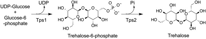
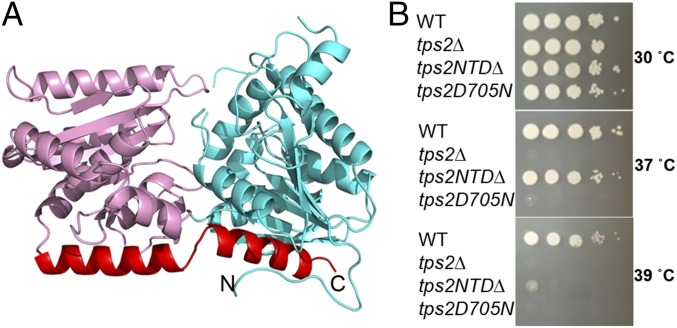
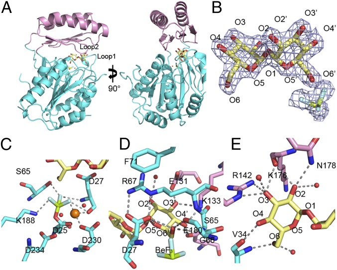
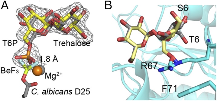
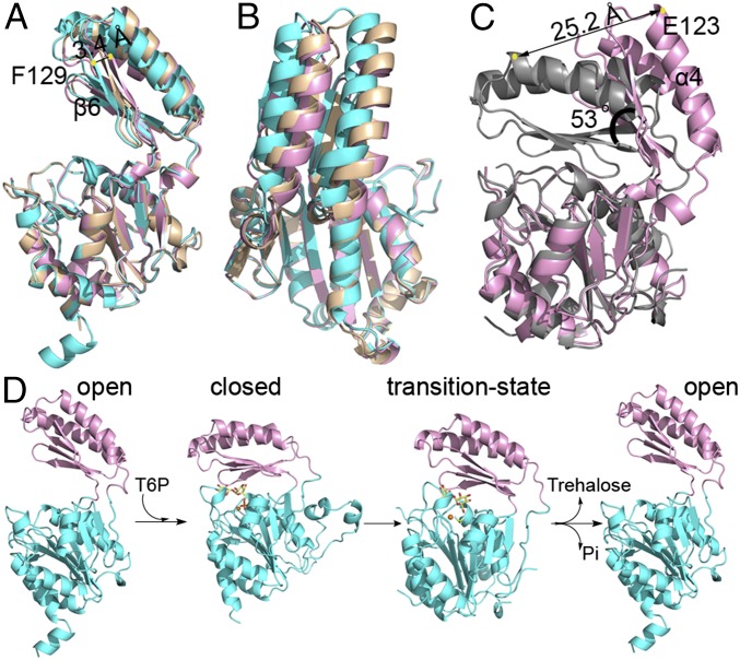
Trehalose is a disaccharide essential for the survival and virulence of pathogenic fungi. The biosynthesis of trehalose requires trehalose-6-phosphate synthase, Tps1, and trehalose-6-phosphate phosphatase, Tps2. Here, we report the structures of the N-terminal domain of Tps2 (Tps2NTD) from Candida albicans, a transition-state complex of the Tps2 C-terminal trehalose-6-phosphate phosphatase domain (Tps2PD) bound to BeF3 and trehalose, and catalytically dead Tps2PD(D24N) from Cryptococcus neoformans bound to trehalose-6-phosphate (T6P). The Tps2NTD closely resembles the structure of Tps1 but lacks any catalytic activity. The Tps2PD-BeF3-trehalose and Tps2PD(D24N)-T6P complex structures reveal a "closed" conformation that is effected by extensive interactions between each trehalose hydroxyl group and residues of the cap and core domains of the protein, thereby providing exquisite substrate specificity. Disruption of any of the direct substrate-protein residue interactions leads to significant or complete loss of phosphatase activity. Notably, the Tps2PD-BeF3-trehalose complex structure captures an aspartyl-BeF3 covalent adduct, which closely mimics the proposed aspartyl-phosphate intermediate of the phosphatase catalytic cycle. Structures of substrate-free Tps2PD reveal an "open" conformation whereby the cap and core domains separate and visualize the striking conformational changes effected by substrate binding and product release and the role of two hinge regions centered at approximately residues 102-103 and 184-188. Significantly, tps2Δ, tps2NTDΔ, and tps2D705N strains are unable to grow at elevated temperatures. Combined, these studies provide a deeper understanding of the substrate recognition and catalytic mechanism of Tps2 and provide a structural basis for the future design of novel antifungal compounds against a target found in three major fungal pathogens.

Keywords: HASDF phosphatase; antifungal inhibitors; pathogenic fungi; trehalose-6-phosphate phosphatase; trehalose-6-phosphate specificity.


Conflict of interest statement

The authors declare no conflict of interest.

Figures

Fig. 1.
Fig. 1.
Enzyme scheme of trehalose biosynthesis. Trehalose is synthesized by the conversion of glucose-6-phosphate and UDP-glucose to trehalose-6-phosphate (T6P) by Tps1 followed by dephosphorylation of T6P by Tps2.
Fig. 2.
Fig. 2.
Structure of the C. albicans Tps2NTD and functional importance of Tps2. (A) Cartoon diagram of the Tps2NTD. The N-terminal and C-terminal Rossmann-fold domains are depicted as ribbons and colored cyan and pink, whereas the C-terminal helix that interacts with both domains is colored red. (B) The phosphatase domain of Tps2 is sufficient to rescue the temperature-sensitive phenotype of tps2∆ in C. neoformans at 37 °C but not at 39 °C, whereas altering a single amino acid residue (D705N), predicted to eliminate Tps2 activity, failed to restore growth at elevated temperatures. The strains were incubated for 3 d.
Fig. 3.
Fig. 3.
Structure of the C. albicans Tps2PD–BeF3–trehalose transition-state complex. (A) Two views of the structure of the Tps2PD–BeF3–trehalose transition-state complex. The core domain and cap domains are shown as cartoons and colored cyan and pink. The trehalose and BeF3 are shown as sticks and the Mg2+ as an orange sphere. (B) A 2Fo-Fc electron density map of the bound trehalose and covalently bound BeF3, shown as light blue mesh and contoured at 1.5 σ. Trehalose is shown as pale yellow sticks. The oxygen atoms are labeled where primed oxygens are found on the glucose ring close to the catalytic residue D25. The beryllium and fluoride are colored olive and light cyan, respectively. (C) View of core-domain residues involved in transition-state stabilization, as depicted by the D25-BeF3 covalent link and Mg2+ ion coordination. The cap-domain residues are shown as atom-colored cyan sticks, and the magnesium ion and waters are depicted as spheres and colored orange and red, respectively. Selected hydrogen bonds are shown by dashed lines. (D) View of residues and solvent involved in binding the catalytic-residue proximal glucose moiety of trehalose. Residues from the cap domain are shown as atom-colored pink sticks and from the core domain as atom-colored cyan sticks. Hydrogen bonds are shown by dashed lines. (E) View of residues and solvent involved in binding the catalytic-residue distal glucose moiety of trehalose. Hydrogen bonds are shown by dashed lines.
Fig. 4.
Fig. 4.
Superpositions reveal key steps in catalysis and substrate specificity of Tps2. (A) View of T6P in the C. neoformans Tps2PD(D24N) closed conformation structure after superposition of the protein onto the Tps2 from the C. albicans Tps2–BeF3–trehalose transition-state complex. Note the significant overlap of the T6P and trehalose sugars. T6P from the C. neoformans Tps2PD structure is shown as atom-colored yellow sticks. A 2Fo-Fc electron density map of T6P bound to the C. neoformans Tps2PD is shown as gray mesh and contoured at 1.5 σ. Trehalose and the D25-BeF3 covalent adduct are shown as atom-colored gray sticks. Mg2+ ions from the C. neoformans and C. albicans Tps2PD structure are shown as orange and gray spheres, respectively. (B) Superposition of the glucose moieties of trehalose and sucrose in the C. albicans Tps2PD–BeF3–trehalose–Mg2+ transition-state complex structure. Trehalose and sucrose are shown as atom-colored gray and yellow sticks, respectively. The 6 position of the phosphorylated glucose and fructose moieties of T6P and S6P is labeled as T6 and S6. The side chains of residues R67 and F71 are shown as cyan- and atom-colored sticks.
Fig. 5.
Fig. 5.
Structures Tps2PD in the open conformation and the catalytic cycle of Tps2. (A) Side view of A. fumigatus Tps2PD structures in the open conformation from three different crystal forms. The Tps2PD structures are depicted as cartoon and displayed after the superposition of their respective core domains. Crystal forms 1, 2, and 3 are colored in cyan, pink, and wheat, respectively. (B) “Top” view of the superposition of the three A. fumigatus Tps2PD structures. (C) Structural superposition of the core domains of A. fumigatus Tps2PD crystal form 2 and the C. albicans Tps2PD transition-state complex. The yellow dots depict the locations of the Cα atom of residue E123 in the closed and open conformations. (D) The Tps2PD catalytic cycle requires conformational changes: Open/apo enzyme ⇒ closed/substrate bound ⇒ transition state/aspartylphosphate formation ⇒ open/product release effected by substrate binding, catalysis, and product release.

References

    1. Brown GD, et al. Hidden killers: Human fungal infections. Sci Transl Med. 2012;4(165):165rv13. - PubMed
    1. Ghannoum MA, Rice LB. Antifungal agents: Mode of action, mechanisms of resistance, and correlation of these mechanisms with bacterial resistance. Clin Microbiol Rev. 1999;12(4):501–517. - PMC - PubMed
    1. Cowen LE. The evolution of fungal drug resistance: Modulating the trajectory from genotype to phenotype. Nat Rev Microbiol. 2008;6(3):187–198. - PubMed
    1. Spaltmann F, Blunck M, Ziegelbauer K. Computer-aided target selection-prioritizing targets for antifungal drug discovery. Drug Discov Today. 1999;4(1):17–26. - PubMed
    1. Gancedo C, Flores CL. The importance of a functional trehalose biosynthetic pathway for the life of yeasts and fungi. FEMS Yeast Res. 2004;4(4-5):351–359. - PubMed

Publication types

MeSH terms

LinkOut - more resources