Review
. 2016 Jun 15;80(3):597-628.
doi: 10.1128/MMBR.00082-15. Print 2016 Sep.

Bacterial Sphingomyelinases and Phospholipases as Virulence Factors

Affiliations
Review

Bacterial Sphingomyelinases and Phospholipases as Virulence Factors

Marietta Flores-Díaz et al. Microbiol Mol Biol Rev. .

Abstract

Bacterial sphingomyelinases and phospholipases are a heterogeneous group of esterases which are usually surface associated or secreted by a wide variety of Gram-positive and Gram-negative bacteria. These enzymes hydrolyze sphingomyelin and glycerophospholipids, respectively, generating products identical to the ones produced by eukaryotic enzymes which play crucial roles in distinct physiological processes, including membrane dynamics, cellular signaling, migration, growth, and death. Several bacterial sphingomyelinases and phospholipases are essential for virulence of extracellular, facultative, or obligate intracellular pathogens, as these enzymes contribute to phagosomal escape or phagosomal maturation avoidance, favoring tissue colonization, infection establishment and progression, or immune response evasion. This work presents a classification proposal for bacterial sphingomyelinases and phospholipases that considers not only their enzymatic activities but also their structural aspects. An overview of the main physiopathological activities is provided for each enzyme type, as are examples in which inactivation of a sphingomyelinase- or a phospholipase-encoding gene impairs the virulence of a pathogen. The identification of sphingomyelinases and phospholipases important for bacterial pathogenesis and the development of inhibitors for these enzymes could generate candidate vaccines and therapeutic agents, which will diminish the impacts of the associated human and animal diseases.


Figures

FIG 1
FIG 1
Roles of different bacterial SMases and PLases in virulence. S. enterica serovar Typhimurium injects SseJ, an SGNH esterase with PLA1 and GCATase activities, via a T3SS into the cytoplasm of the host cell. Once in the cytosol, SseJ binds to RhoA GTPase, triggering its GCATase activity, which increases the vacuole surface. R. typhi produces two patatin-like PLA2s, Pat1 and Pat2, which are secreted during host intracellular growth and help phagosome escape. S. pyogenes SlaA is a class I-like PLA2 that enters host epithelial cells in an actin-dependent manner and plays an important role in pathogen adhesion and cytotoxicity. P. aeruginosa injects ExoU, a patatin-like PLA2, through a T3SS into the cytoplasm of the host cell. Once translocated, ExoU becomes activated and acts toward several plasma membrane substrates, leading to cytoskeletal collapse and cellular necrosis. ExoU also activates several signaling pathways, such as the arachidonic acid cascade and a PAF receptor–NF-κB pathway that leads to inflammatory mediator production. L. monocytogenes produces the PI-PLC PlcA and the Zn2+ metalloPLC PlcB, which are required for bacterial escape into the cytosol from the single-membrane primary pathogen-containing vacuole, triggering preautophagosomal structure stalling, favoring bacterial escape from the host autophagic defense. L. monocytogenes PlcB also contributes to bacterial escape into the cytoplasm. Both Bacillus spp. and S. aureus secrete a SMase C that plays an important role in virulence by increasing Cer, which changes the physical properties of the membrane and could also participate in signal transduction leading to cell death. These two bacteria also produce PI-PLC, which can facilitate signal transduction and removal of GPI-anchored proteins from plasma membranes. C. perfringens alpha-toxin, a Zn2+ metalloPLC, is required for this bacterium's escape from the macrophage phagosome in early stages of infection. At high concentrations, this toxin disrupts the plasma membrane, causing cell lysis, but at low concentrations if internalized by the target cell it cleaves PC and SM at the membranes of the endolysosomal compartment, generating DAG and Cer, which trigger signaling pathways that lead to ROS production and cell death. N. gonorrhoeae secretes a PLD named NgPLD that by both generation of PA and Akt binding drives cell surface recruitment of CR3, membrane ruffling, and bacteria engulfment, modulating host cell signaling events required for bacterial invasion.
FIG 2
FIG 2
SMase cleavage sites in sphingomyelin and cellular activities of the reaction products. SMase Cs cleave SM, generating Cer and phosphorylcholine, while SMase Ds generate Cer-1-phosphate and choline. SMase Ds also have intrinsic lysophospholipase D activities and act on lysophosphatidylcholine to generate the signaling molecule LPA (see also Fig. 5).
FIG 3
FIG 3
Structure of S. aureus beta-toxin (PDB ID 3I5V). The structure is shown as a beige cartoon, with catalytically important residues and bound diacyl glycerol shown as beige and gray sticks, respectively. Mg2+ ions from B. cereus SMase C (PDB ID 2DDT) are superimposed and shown as light blue spheres, and phosphate from L. ivanovii Smase C (PDB ID 1ZWX) is shown as sticks.
FIG 4
FIG 4
Molecular model of C. pseudotuberculosis Smase D. (A) Model based on the structure of the spider Loxosceles laeta Smase D (PDB ID 1XX1), shown as a blue cartoon, with the torus-capping C-terminal helix and strand in pink. The catalytically essential Mg2+ ion is shown as a green sphere, with the completely conserved coordinating aspartates and glutamates shown as sticks. A sulfate ion also coordinating the Mg2+ and likely an analogue for the sphingomyelin phosphate moiety is shown as sticks. (B) Model similar to that in panel A, but from the viewpoint of looking down into the active site, with a semitransparent electrostatic surface drawn highlighting the hydrophobic loops (white) and negatively charged center (red) that forms the active site.
FIG 5
FIG 5
PLase cleavage sites in phosphatidylcholine and cellular activities of the reaction products. PLA1s remove the fatty acid at the sn-1 position of the glycerol moiety, while PLA2s remove it at the sn-2 position, generating cellular mediators like lysophosphatidylcholine and arachidonic acid. PLBs hydrolyze both acyl groups from the glycerophospholipid and also exhibit LPLA activity. PLCs hydrolyze the glycerol-oriented phosphodiester bond, releasing DAG and a phosphorylated head group [e.g., IP3, acting on PI(4,5)P2, or choline phosphate acting on PC]. PLDs cleave the alcohol-oriented phosphodiester bond, releasing the head group (e.g., choline or inositol) and generating LPA. (Based on data from reference .)
FIG 6
FIG 6
Molecular model of the Helicobacter pylori PldA1 dimer. (A) The model based on E. coli OMPLA (PDB ID 1QD6), shown as a pink and green cartoon. The inhibitor hexadecanoylsulphoic acid is shown as sticks at the dimer interface, and the Ser-His-Gln catalytic triad is shown as sticks and labeled. The Ca2+ ion that enhances activity by influencing active-site electrostatics is shown as a blue sphere, with its coordinating main-chain carbonyl ions drawn as sticks. (B) Increased magnification image of the binding pocket lined with hydrophobic residues at the dimer interface. Colors are as for panel A, but with all residues drawn as sticks and with a semitransparent molecular surface to highlight the large pocket that can accommodate diverse lipid tails.
FIG 7
FIG 7
Molecular model of M. bovis PLB. The model is based on P. aeruginosa EstA (PDB ID 3KVN), and the active transporter is drawn as a gold cartoon, with the conserved blocks of residues associated with acyl hydrolase activity shown in red and the residues of the catalytic motif shown as sticks.
FIG 8
FIG 8
Structures of P. aeruginosa ExoU and its chaperone, SpcU (PDB ID 3TU3), shown as a cartoon. SpcU is shown in white, the ExoU chaperone binding domain is in gold, the PLA2 domain is green, and the membrane localization domain is shown in pink and purple. Catalytically important residues are shown as yellow sticks, and the ubiquitylable Lys178 is shown as a space-filling representation.
FIG 9
FIG 9
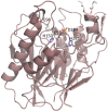
Structure of L. monocytogenes PI-PLC (PDB ID 2PLC), shown as a cartoon. Bound myo-inositol is shown as gray sticks. General base His45, general acid His93, and phosphate-interacting residue Arg84, as well as residues interacting with the PI head group. are shown as sticks and labeled.
FIG 10
FIG 10
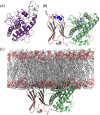
Structures of Zn2+ metalloPLCs from B. cereus and C. perfringens, similarly oriented. (A) B. cereus PLC (PDB ID 1P6D) is shown as a purple cartoon, with inserted hairpin in white. Active-site Zn2+ ions are shown as lighter-colored spheres. The bound PC analogue is shown by white sticks. (B) C. perfringens alpha-toxin (PDB ID 1CA1) with the N terminal (homologous to B. cereus PLC) in green and membrane-binding C terminus in pink. Zn2+ (violet) and Ca2+ (blue) ions are shown as spheres, and coordinating residues are shown as sticks. (C) The alpha-toxin shown in panel B, modeled bound to mixed PC/cholesterol membrane, highlighting how the B. cereus hairpin would impinge into the membrane.
FIG 11
FIG 11
General scheme of the events triggered by C. perfringens alpha-toxin during gas gangrene. The pleiotropic effects of C. perfringens alpha-toxin in diverse cell types contribute to reducing vascular perfusion and causing tissue damage, which enhance the conditions for bacterial growth and lead to shock and multiple organ failure.
FIG 12
FIG 12
Molecular model of P. aeruginosa PlcH. The model is based on P. aeruginosa ApcA (PDB ID 2D1G) and is shown as a gold cartoon, with active-site residues conserved between the two enzymes shown as sticks. The metal ion found in the ApcA structure is shown as a blue sphere, and the phosphate analogue is shown as white sticks. The majority of the conserved residues interact with either the metal ion or the phosphate analogue, with the exception of the likely nucleophile, Thr178 (labeled).
FIG 13
FIG 13
Molecular model of Acinetobacter baumanii PLD1. The model is based on the structure of the Streptomyces PLD (PDB ID 1F0I) and is shown as a cartoon. The N-terminal domain is shaded green, and the C terminal is pink. Residues at the active site and part of the PLD motif are shown as sticks, and the covalently bound reaction intermediate is shown as white sticks at the interface between the domains. Note the symmetrically positioned motif residues in each homologous domain.
None
None
None
None
None

References

    1. Lingwood D, Simons K. 2010. Lipid rafts as a membrane-organizing principle. Science 327:46–50. doi: 10.1126/science.1174621. - DOI - PubMed
    1. van Meer G, Voelker DR, Feigenson GW. 2008. Membrane lipids: where they are and how they behave. Nat Rev Mol Cell Biol 9:112–124. doi: 10.1038/nrm2330. - DOI - PMC - PubMed
    1. Goñi FM, Alonso A. 1999. Structure and functional properties of diacylglycerols in membranes. Prog Lipid Res 38:1–48. doi: 10.1016/S0163-7827(98)00021-6. - DOI - PubMed
    1. Castro BM, Prieto M, Silva LC. 2014. Ceramide: a simple sphingolipid with unique biophysical properties. Prog Lipid Res 54:53–67. doi: 10.1016/j.plipres.2014.01.004. - DOI - PubMed
    1. Stancevic B, Kolesnick R. 2010. Ceramide-rich platforms in transmembrane signaling. FEBS Lett 584:1728–1740. doi: 10.1016/j.febslet.2010.02.026. - DOI - PMC - PubMed

Publication types

MeSH terms

LinkOut - more resources