Review
. 2016 Jun 15;80(3):629-61.
doi: 10.1128/MMBR.00078-15. Print 2016 Sep.

Klebsiella pneumoniae: Going on the Offense with a Strong Defense

Affiliations
Review

Klebsiella pneumoniae: Going on the Offense with a Strong Defense

Michelle K Paczosa et al. Microbiol Mol Biol Rev. .

Abstract

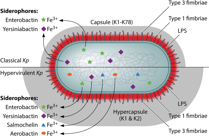
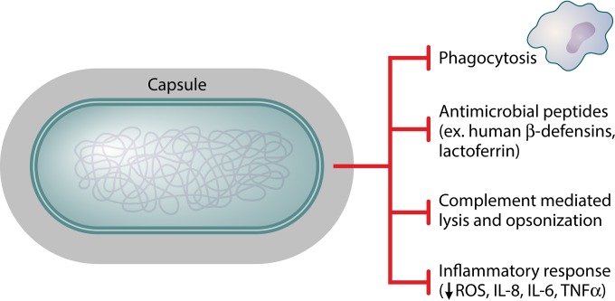
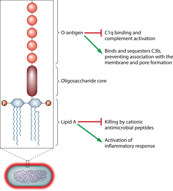
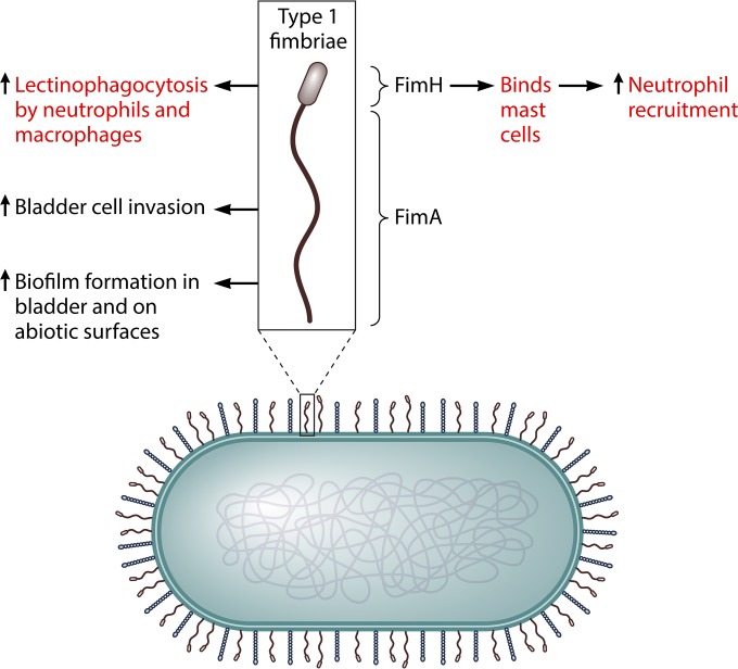
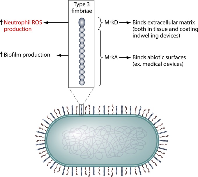
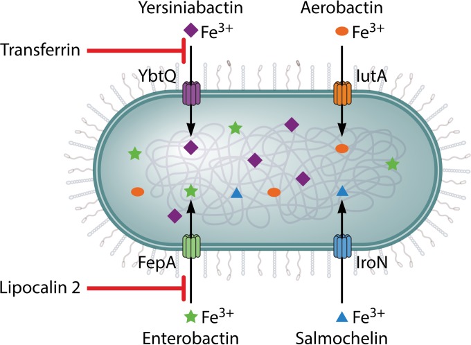
Klebsiella pneumoniae causes a wide range of infections, including pneumonias, urinary tract infections, bacteremias, and liver abscesses. Historically, K. pneumoniae has caused serious infection primarily in immunocompromised individuals, but the recent emergence and spread of hypervirulent strains have broadened the number of people susceptible to infections to include those who are healthy and immunosufficient. Furthermore, K. pneumoniae strains have become increasingly resistant to antibiotics, rendering infection by these strains very challenging to treat. The emergence of hypervirulent and antibiotic-resistant strains has driven a number of recent studies. Work has described the worldwide spread of one drug-resistant strain and a host defense axis, interleukin-17 (IL-17), that is important for controlling infection. Four factors, capsule, lipopolysaccharide, fimbriae, and siderophores, have been well studied and are important for virulence in at least one infection model. Several other factors have been less well characterized but are also important in at least one infection model. However, there is a significant amount of heterogeneity in K. pneumoniae strains, and not every factor plays the same critical role in all virulent Klebsiella strains. Recent studies have identified additional K. pneumoniae virulence factors and led to more insights about factors important for the growth of this pathogen at a variety of tissue sites. Many of these genes encode proteins that function in metabolism and the regulation of transcription. However, much work is left to be done in characterizing these newly discovered factors, understanding how infections differ between healthy and immunocompromised patients, and identifying attractive bacterial or host targets for treating these infections.


Figures

FIG 1
FIG 1
Four well-characterized virulence factors in classical and hypervirulent K. pneumoniae (Kp) strains. There are four well-characterized virulence factors for pathogenic K. pneumoniae: capsule, LPS, fimbriae (type 1 and type 3), and siderophores. Capsule is an extracellular polysaccharide matrix that envelops the bacteria. Classical K. pneumoniae strains produce a capsule that can be of any of the serotypes K1 to K78; K1 and K2 are associated with increased pathogenicity. HV strains make a hypercapsule, which amplifies the production of capsular material, resulting in a relatively larger capsule, and are predominantly of the K1 serotype, while the remaining strains are of serotype K2. LPS, an integral part of the outer leaflet of the outer membrane, is produced by both classical and HV K. pneumoniae strains and can be of O-antigen serotypes 1 to 9 (O1 to -9). Both types of K. pneumoniae make membrane-bound adhesive structures, type 1 and type 3 fimbriae, and secrete iron-scavenging siderophores. Of the siderophores, enterobactin is made by almost all strains, and yersiniabactin is made by approximately half of classical and almost all HV strains. Salmochelin and aerobactin are rarely produced by classical strains but are typically secreted by HV strains, with aerobactin being the most highly expressed of the siderophores.
FIG 2
FIG 2
Role of capsule in K. pneumoniae virulence. A number of different functions for capsule have been delineated for K. pneumoniae virulence. First, capsule prevents phagocytosis and opsonophagocytosis of the bacteria by immune cells. Second, it hinders the bactericidal action of antimicrobial peptides such as human beta defensins 1 to 3 and lactoferrin by binding these molecules distal from the outer membrane. Third, it blocks complement components, such as C3, from interacting with the membrane, thus preventing complement-mediated lysis and opsonization. Finally, it averts the fulminant activation of the immune response, as measured by decreased reactive oxygen species (ROS), IL-8, IL-6, and TNF-α production, by assisting in the activation of a NOD-dependent pathway and shielding LPS from recognition by immune cell receptors.
FIG 3
FIG 3
Role of lipopolysaccharide in K. pneumoniae virulence. LPS is composed of three major subunits: lipid A, an oligosaccharide core, and O antigen. Lipid A inserts into the bacterial membrane and is a potentially potent activator of inflammation. K. pneumoniae may modify its lipid A to make it less inflammatory during infection, and lipid A may also protect against the bactericidal action of cationic antimicrobial peptides. O antigen is the outermost subunit of LPS. It has important roles in protecting against complement, including preventing C1q binding to bacteria, which inhibits subsequent activation of the complement pathway, as well as binding C3b away from the outer bacterial membrane and, thus, abrogating bacterial lysis by the complement membrane attack complex.
FIG 4
FIG 4
Functions of type 1 fimbriae during K. pneumoniae infection and biofilm formation. Type 1 fimbriae are filamentous, membrane-bound, adhesive structures composed primarily of FimA subunits, with the FimH subunit on the tip. These fimbriae have a role in bladder cell invasion by K. pneumoniae as well as biofilm formation in the bladder and on abiotic surfaces. However, type 1 fimbriae may be a negative influence on K. pneumoniae virulence in vivo in a few ways. First, type 1 fimbriae amplify lectinophagocytosis of K. pneumoniae by macrophages and neutrophils. Second, the FimH subunit increases binding to immune cells such as mast cells, leading to increased immune cell activation and subsequent recruitment of neutrophils, which likely increases K. pneumoniae clearance.
FIG 5
FIG 5
Functions of type 3 fimbriae during K. pneumoniae infection and biofilm formation. Type 3 fimbriae are helix-like, membrane-bound, adhesive structures on the surface of K. pneumoniae. They are composed primarily of MrkA subunits, with the MrkD subunit on the tip. Type 3 fimbriae have been found to be necessary for K. pneumoniae biofilm production and binding to medical devices. MrkD has specifically been found to bind extracellular matrix, such as that exposed on damaged tissues and coating indwelling devices, while MrkA binds abiotic surfaces, such as medical devices both prior to insertion into patients and after insertion when coated with host matrix. Type 3 fimbriae have been shown to have a possibly detrimental role, as their presence on K. pneumoniae increases reactive oxygen species (ROS) production by neutrophils.
FIG 6
FIG 6
Siderophore production and roles in virulence in K. pneumoniae. The ability to acquire iron in an iron-poor environment during infection is necessary for K. pneumoniae pathogenesis. Therefore, bacteria secrete proteins with a high affinity for iron, called siderophores. K. pneumoniae strains have been found to produce one or more of the following siderophores: enterobactin, salmochelin, yersiniabactin, and aerobactin. Enterobactin is the primary siderophore used by K. pneumoniae, although it is inhibited by the host molecule lipocalin-2. Salmochelin is a c-glucosylated form of enterobactin that can no longer be inhibited by lipocalin-2. Yersiniabactin and aerobactin are structurally distinct from enterobactin and salmochelin. Neither siderophore can be inhibited by lipocalin-2, but yersiniabactin functionality is reduced in the presence of the host molecule transferrin. The production of a number of different siderophores may allow K. pneumoniae to colonize and disseminate to a number of different sites within the host, with niche-specific roles for each siderophore. FepA, IroN, YbtQ, and IutA serve as transporters specific to their corresponding siderophores of enterobactin, salmochelin, yersiniabactin, and aerobactin, respectively.

Similar articles

Cited by

References

    1. Friedlander C. 1882. Uber die scizomyceten bei der acuten fibrosen pneumonie. Arch Pathol Anat Physiol Klin Med 87:319–324.
    1. Merino S, Camprubi S, Alberti S, Benedi VJ, Tomas JM. 1992. Mechanisms of Klebsiella pneumoniae resistance to complement-mediated killing. Infect Immun 60:2529–2535. - PMC - PubMed
    1. Bagley ST. 1985. Habitat association of Klebsiella species. Infect Control 6:52–58. - PubMed
    1. Rock C, Thom KA, Masnick M, Johnson JK, Harris AD, Morgan DJ. 2014. Frequency of Klebsiella pneumoniae carbapenemase (KPC)-producing and non-KPC-producing Klebsiella species contamination of healthcare workers and the environment. Infect Control Hosp Epidemiol 35:426–429. doi:10.1086/675598. - DOI - PMC - PubMed
    1. Dao TT, Liebenthal D, Tran TK, Ngoc Thi Vu B, Ngoc Thi Nguyen D, Thi Tran HK, Thi Nguyen CK, Thi Vu HL, Fox A, Horby P, Van Nguyen K, Wertheim HFL. 2014. Klebsiella pneumoniae oropharyngeal carriage in rural and urban Vietnam and the effect of alcohol consumption. PLoS One 9:e91999. doi:10.1371/journal.pone.0091999. - DOI - PMC - PubMed

MeSH terms

LinkOut - more resources