. 2016 Sep;15(9):2187-97.
doi: 10.1158/1535-7163.MCT-15-0427. Epub 2016 Jun 15.

Periostin (POSTN) Regulates Tumor Resistance to Antiangiogenic Therapy in Glioma Models

Affiliations

Periostin (POSTN) Regulates Tumor Resistance to Antiangiogenic Therapy in Glioma Models

Soon Young Park et al. Mol Cancer Ther. 2016 Sep.

Abstract

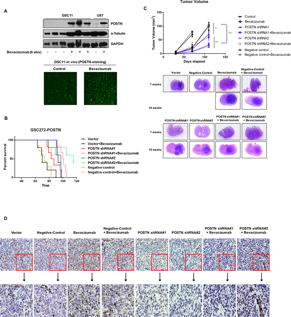
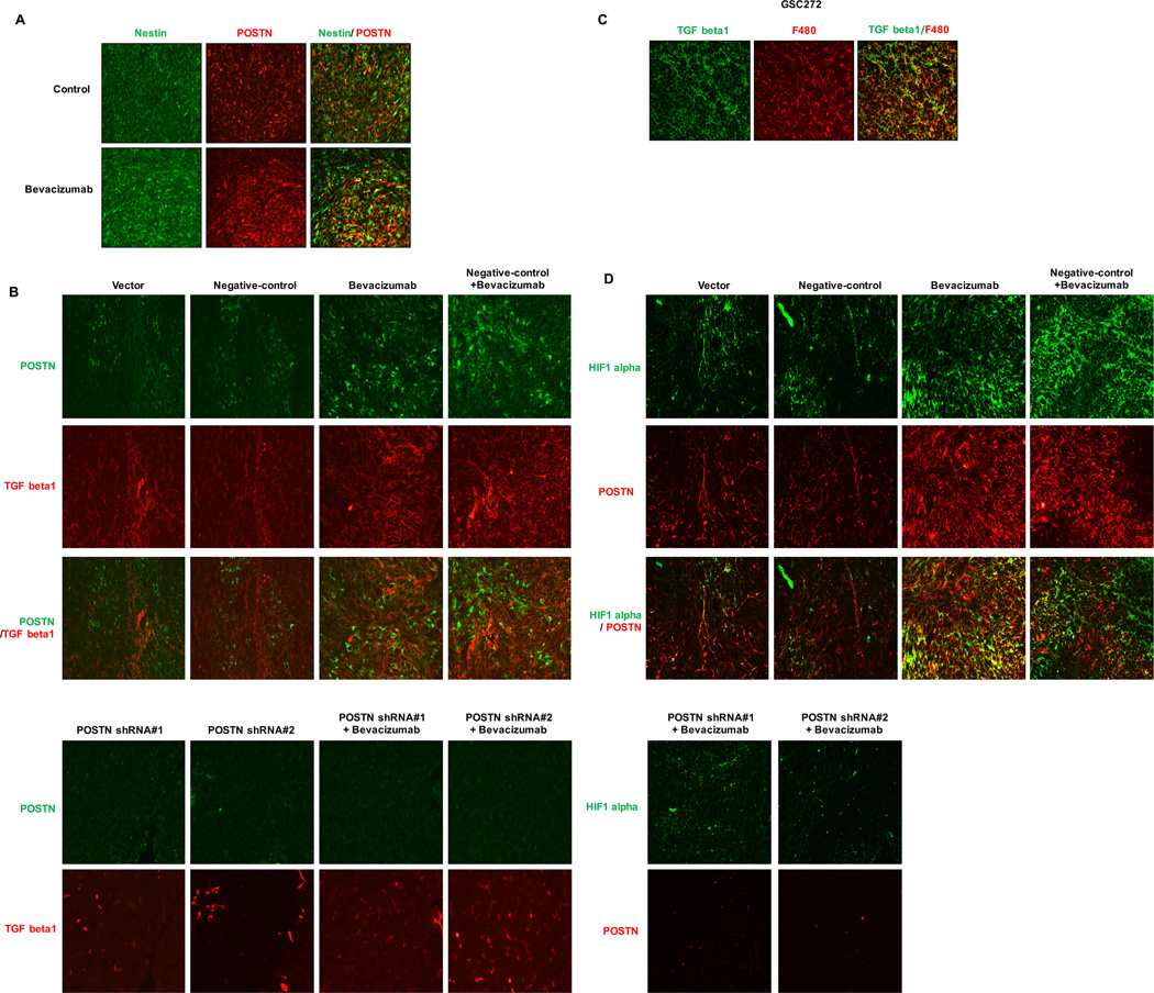
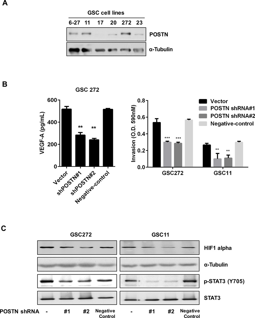
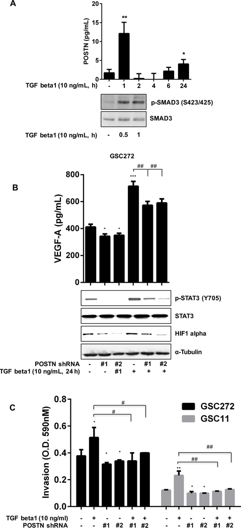
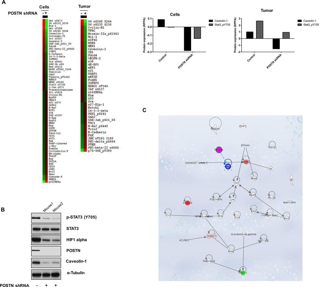
Periostin (POSTN) interacts with multiple integrins to coordinate a variety of cellular processes, including epithelial-to-mesenchymal transition (EMT) and cell migration. In our previous study, anti-VEGF-A therapy was associated with resistance and EMT. This study sought to determine the role of POSTN in the resistance of glioma stem cells (GSC) to antiangiogenic therapy. In mouse xenograft models of human glioma, POSTN expression was associated with acquired resistance to anti-VEGF-A therapy and had a synergistic effect with bevacizumab in prolonging survival and decreasing tumor volume. Resistance to anti-VEGF-A therapy regulated by POSTN was associated with increased expression of TGFβ1 and hypoxia-inducible factor-1α (HIF1α) in GSCs. At the molecular level, POSTN regulated invasion and expression of EMT (caveolin-1) and angiogenesis-related genes (HIF1α and VEGF-A) through activation of STAT3. Moreover, recombinant POSTN increased GSC invasion. Collectively, our findings suggest that POSTN plays an important role in glioma invasion and resistance to antiangiogenic therapy. Mol Cancer Ther; 15(9); 2187-97. ©2016 AACR.


Conflict of interest statement

of Potential Conflicts of Interest: John de Groot reports serving on an advisory board for Genentech/Roche. The other authors declare no conflict of interest.

Figures

Figure 1
Figure 1
Knockdown of POSTN expression prolongs survival and decreases tumor volume in glioma mouse models. (A) GSC11 and U87 cells were intracranially transplanted into the brains of mice (5 × 10⁵ cells/mouse). Afterward, bevacizumab was administered via intraperitoneal injection. Proteins were collected from tumors (control group, at 4 weeks; bevacizumab group, at 6 weeks), and POSTN expression was detected by Western blot analysis. POSTN expression (green) was detected on frozen sections of mouse brain tumors by immunofluorescent staining. (B) The effects of POSTN knockdown and bevacizumab treatment on GBM growth and survival were examined in a mouse intracranial xenograft model. GSC272 cells were infected with a lentivirus containing vector shRNA, POSTN shRNA#1, POSTN shRNA#2 or negative control shRNA and then intracranially transplanted into the brains of immunocompromised mice (5 × 10⁵ cells/mouse). Tumors derived from POSTN-knockdown GSCs, with or without bevacizumab treatment, were harvested simultaneously and examined to determine the impact of disruption of POSTN expression. Survival of the study mice was estimated by the Kaplan-Meier method. In the GSC272 group, the following were compared: control versus treatment with bevacizumab (P = 0.001), control versus POSTN knockdown (#1: P = 0.05; #2: P = 0.013), POSTN knockdown versus POSTN knockdown plus treatment with bevacizumab (#1: P = 0.01; #2: P = 0.002), treatment with bevacizumab versus POSTN knockdown plus treatment with bevacizumab (#1: P = 0.044; #2: P = 0.002). (C) Murine brains harvested on day 30, 70, or 110 after transplantation of parental GSCs or POSTN-knockdown GSCs with or without treatment with bevacizumab were stained with H&E. At 70 days, treatment with bevacizumab and knockdown of POSTN expression resulted in smaller tumors than those in control mice (*P < 0.05). At 110 days, the combination resulted in smaller tumors than those in the bevacizumab-treated (***P < 0.001) and POSTN-knockdown (**P < 0.01) groups. (D) Immunohistochemical stains of GSC272 tumor samples for POSTN. POSTN-positive cells stained in nuclei.
Figure 2
Figure 2
Treatment with bevacizumab increases the expression of HIF1 alpha and TGF beta1 via POSTN. Immunofluorescent stains are shown for POSTN and the GSC markers nestin (A), TGF beta1 (B), and HIF1 alpha (C) in GSC272 mouse glioma xenografts with or without POSTN knockdown and/or treatment with bevacizumab. (A) Expression of POSTN (red) and of nestin (green) overlaps. (B) TGF beta1 (red) and POSTN (green) were expressed at different sites. (C) TGF beta1 (green) was expressed in F4/80-positive cells (red). (D) Immunofluorescent stains for POSTN (red) in relation to hypoxia marked by HIF1 alpha staining (green).
Figure 3
Figure 3
POSTN regulates invasiveness and VEGF-A expression in GSCs. (A) GSC cell lysates were collected and analyzed for POSTN expression by Western blot. (B and C) GSC11 and GSC272 cells were infected with a lentivirus containing a control vector, POSTN shRNA or negative-control. (B) Expression of VEGF-A in the cells was measured by a VEGF-A ELISA kit. The invasiveness of GSCs (vector-, POSTN shRNA-infected or negative-control) was assessed by an invasion assay for 18 h. **P < 0.01, ***P < 0.001 versus control. (C) Expression of HIF1 alpha and STAT3 in the GSCs was detected by Western blotting.
Figure 4
Figure 4
TGF beta1 increases pSTAT3, HIF1 alpha, and VEGF-A expression in and invasiveness of GSCs. (A) GSC272 cells were seeded in 6-well plates for 24 h and treated with TGF beta1 (in medium without EGF or bFGF) for the indicated times. Secretion of POSTN and expression of SMAD3 in GSC272 cells were assessed by ELISA and Western blot analysis, respectively. *P < 0.05, **P < 0.01 versus control. (B) GSC272 cells infected with vector shRNA or POSTN shRNA were treated with TGF beta1 for 24 h; expression of VEGF-A was determined by ELISA and expression of p-STAT3, STAT3, and HIF1 alpha was determined by Western blot. *P < 0.05, ***P < 0.001 versus control; ##P < 0.01 versus TGF beta1. (C) GSC272 cells infected with a vector or POSTN shRNA were added to the upper chambers of a transwell insert (2 × 10⁴ cells/well), and medium containing recombinant TGF beta1 was added to the lower chambers; the plate was incubated for 18 h at 37°C. *P < 0.05, **P < 0.01 versus control; #P < 0.05, ##P < 0.01 versus TGF beta1.
Figure 5
Figure 5
Recombinant POSTN and TGF beta1 increase GSC invasiveness via integrin β1. (A) Expression of integrin β1 and integrin β3 proteins was measured in several GSC lines by Western blotting. (B) GSCs (2 × 10⁴ cells/well) were rotated with antibody to integrin β1 or β3 for 1 h at room temperature, and cells were added to the upper chambers of a transwell insert plate. Medium containing recombinant POSTN or TGF beta1 was added to the lower chambers, and the plate was incubated for 18 h at 37°C. **P < 0.01, ***P < 0.001 versus control; ##P < 0.01, ###P < 0.001 versus POSTN or TGF beta1. (C) Vector- and POSTN shRNA-infected GSC272 cells were treated with 2 µg/mL recombinant POSTN for 24 h. Phosphorylation of STAT3 and expression of POSTN and caveolin-1 in the cells were assessed by Western blotting.
Figure 6
Figure 6
Expression of proteins related to POSTN were quantified in GSCs and GSC-derived mouse tumors by RPPA. (A) Expression of various proteins was determined in GSC272 cells (vector- or POSTN shRNA-infected) and murine glioma samples derived from GSC272 (vector- or POSTN shRNA-infected) at 12 weeks after implantation. The protein expression levels in POSTN shRNA-infected cells and tumors that differed at least 1.3-fold (GSC272 cells) or 0.7-fold (murine glioma samples) from controls were selected for hierarchical clustering analysis. The red and green colors reflect relatively high and low expression, respectively. (B) Western blot analysis detected proteins expression in murine GSC272 glioma samples with and without POSTN knockdown. (C) Ingenuity Pathway Analysis examined the intercellular signaling interaction network identified by in vitro and in vivo RPPA results.

References

    1. Arrillaga-Romany I, Reardon DA, Wen PY. Current status of antiangiogenic therapies for glioblastomas. Expert Opin Investig Drugs. 2014;23:199–210. - PubMed
    1. Liu LX, Lu H, Luo Y, Date T, Belanger AJ, Vincent KA, et al. Stabilization of vascular endothelial growth factor mRNA by hypoxia-inducible factor 1. Biochem Biophys Res Commun. 2002;291:908–914. - PubMed
    1. Bao S, Wu Q, Sathornsumetee S, Hao Y, Li Z, Hjelmeland AB, et al. Stem cell-like glioma cells promote tumor angiogenesis through vascular endothelial growth factor. Cancer Res. 2006;66:7843–7848. - PubMed
    1. Takano S, Yoshii Y, Kondo S, Suzuki H, Maruno T, Shirai S, et al. Concentration of vascular endothelial growth factor in the serum and tumor tissue of brain tumor patients. Cancer Res. 1996;56:2185–2190. - PubMed
    1. Soda Y, Myskiw C, Rommel A, Verma IM. Mechanisms of neovascularization and resistance to anti-angiogenic therapies in glioblastoma multiforme. J Mol Med. 2013;91:439–448. - PMC - PubMed

Publication types

MeSH terms