Case Reports
. 2015 Nov 6;5(1):327.
doi: 10.2484/rcr.v5i1.327. eCollection 2010.

Uterine didelphys associated with obstructed hemivagina and ipsilateral renal anomaly (OHVIRA) syndrome

Case Reports

Uterine didelphys associated with obstructed hemivagina and ipsilateral renal anomaly (OHVIRA) syndrome

Boram Han et al. Radiol Case Rep. .

Abstract

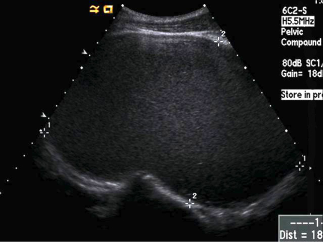
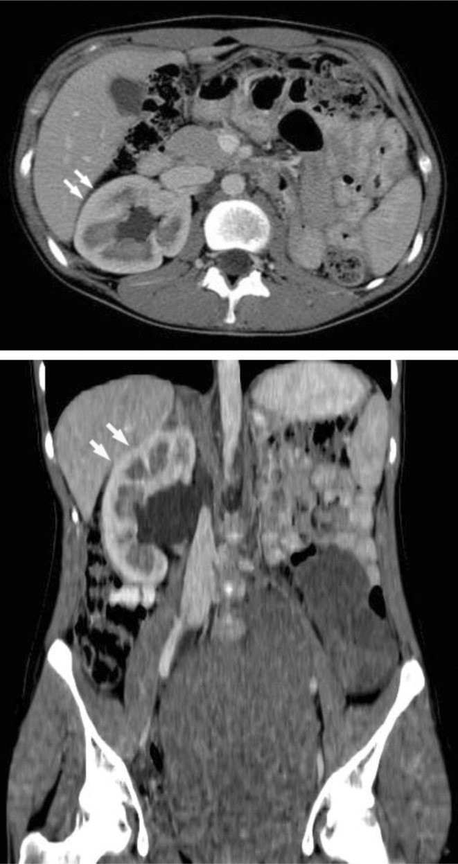
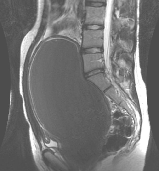
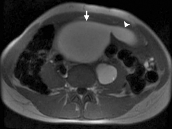
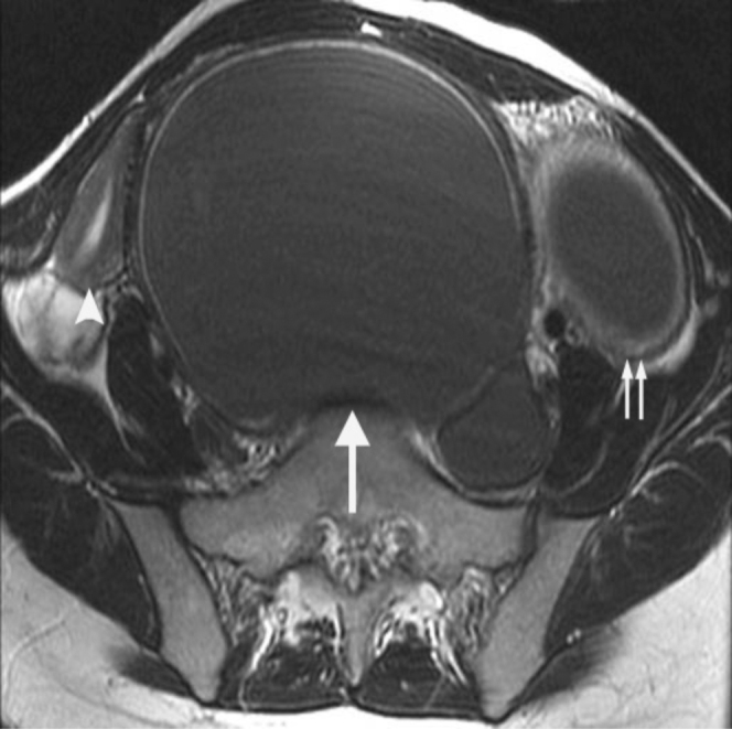
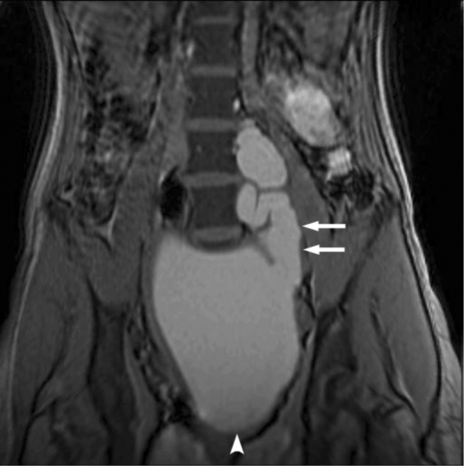
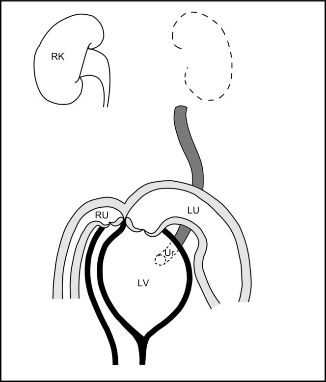
Obstructed hemivagina and ipsilateral renal anomaly (OHVIRA) syndrome is a rare complex of structural abnormalities of the female urogenital tract. A 17-year-old girl with uterine didelphys associated with OHVIRA syndrome presented with progressive development of cyclic lower abdominal discomfort and a large abdominopelvic mass. We describe the findings from ultrasound, computed tomography (CT), and magnetic resonance imaging (MRI), the first case report of this syndrome to examine all three different imaging modalities in a single patient. We also review the literature on OHVIRA syndrome and discuss important considerations relevant to radiologists and other clinicians.

Keywords: CT, computed tomography; MRI, magnetic resonance imaging.


Figures

Figure 1
Figure 1
17-year-old girl with uterine didelphys associated with OHVIRA syndrome. Longitudinal transabdominal sonographic image through the pelvis demonstrates a markedly dilated vagina, approximately 18×11 cm, containing echogenic material consistent with blood product.
Figure 2
Figure 2
17-year-old girl with uterine didelphys associated with OHVIRA syndrome. Coronal-reformatted, contrast-enhanced CT image demonstrates a blood-filled, markedly dilated vagina (large single arrow), normal right hemiuterine horn (arrowhead), and distended left hemiuterine horn (small paired arrows) with an attenuation of 70 Hounsfield units, suggesting the presence of blood in it.
Figure 3
Figure 3
17-year-old girl with uterine didelphys associated with OHVIRA syndrome. Axial contrast-enhanced CT image (top) and coronal-reformatted, contrast-enhanced CT image (above) demonstrate a mildly hypertrophied right kidney (arrows) and an absent left kidney.
Figure 4
Figure 4
17-year-old girl with uterine didelphys associated with OHVIRA syndrome. Sagittal T2-weighted MR image demonstrates a dilated hemivagina with low T2-signal-intensity contents and corresponding high T1-signal intensity consistent with blood product.
Figure 5
Figure 5
17-year-old girl with uterine didelphys associated with OHVIRA syndrome. Axial T1-weighted MR image demonstrates a left hydrosalpinx (arrowhead) containing high T1-signal-intensity contents consistent with blood products, with an axial diameter of 5.2 cm, originating from a dilated hemivagina (arrow).
Figure 6
Figure 6
17-year-old girl with uterine didelphys associated with OHVIRA syndrome. Axial T2-weighted MR image demonstrates a dilated vagina with low T2-signal-intensity contents that had corresponding high T1-signal intensity consistent with blood products (large single arrow). Normal-appearing right uterine horn (arrowhead) and dilated left uterine horn with low T2-signal-intensity blood products (small paired arrows) also appear.
Figure 7
Figure 7
17-year-old girl with uterine didelphys associated with OHVIRA syndrome. Coronal T1-weighted MR image demonstrates an ectopic insertion of a dilated ureter (paired arrows), containing high T1-signal-intensity contents consistent with refluxed blood products, into a dilated left vaginal canal (arrowhead).
Figure 8
Figure 8
17-year-old girl with uterine didelphys associated with OHVIRA syndrome. Diagrammatic representation of the anatomy of this patient. LV: obstructed left hemivagina dilated with blood product. RU: nondilated right uterine horn. LU: left uterine horn dilated with blood product. Ur: ectopic ureter inserting into the left hemivagina. RK: right kidney.

References

    1. Shavell VI, Montgomery SE, Johnson SC, Diamond MP, Berman JM. Complete septate uterus, obstructed hemivagina, and ipsilateral renal anomaly: pregnancy course complicated by a rare urogenital anomaly. Arch Gynecol Obstet. 2009 Sep;280(3):449–452. [PubMed] - PubMed
    1. Smith NA, Laufer MR. Obstructed hemivagina and ipsilateral renal anomaly (OHVIRA) syndrome: management and follow-up. Fertil Steril. 2007 Apr;87(4):918–922. [PubMed] - PubMed
    1. Gholoum S, Puligandla PS, Hui T, Su W, Quiros E, Laberge JM. Management and outcome of patients with combined vaginal septum, bifid uterus, and ipsilateral renal agenesis (Herlyn-Werner-Wunderlich syndrome) J Pediatr Surg. 2006 May;41(5):987–992. [PubMed] - PubMed
    1. Herlyn U, Werner H. [Simultaneous occurrence of an open Gartner-duct cyst, a homolateral aplasia of the kidney and a double uterus as a typical syndrome of abnormalities] Geburtshilfe Frauenheilkd. 1971 Apr;31(4):340–347. [PubMed] - PubMed
    1. Wunderlich M. [Unusual form of genital malformation with aplasia of the right kidney] Zentralbl Gynakol. 1976;98(9):559–562. [PubMed] - PubMed

Publication types

LinkOut - more resources