Case Reports
. 2015 Nov 6;5(1):332.
doi: 10.2484/rcr.v5i1.332. eCollection 2010.

Recurrent cholecystitis and cholelithiasis in a gallbladder remnant 14 years after a converted cholecystectomy

Case Reports

Recurrent cholecystitis and cholelithiasis in a gallbladder remnant 14 years after a converted cholecystectomy

Sean K Calhoun et al. Radiol Case Rep. .

Abstract

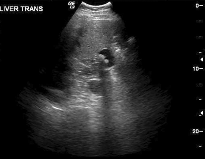
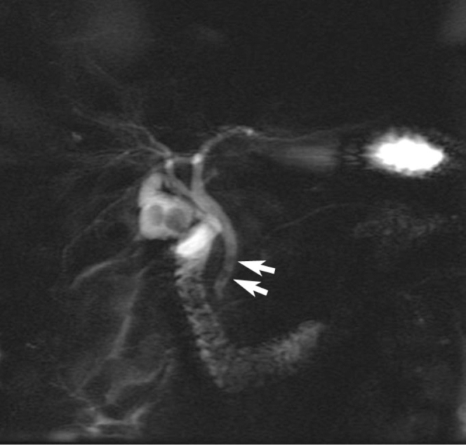
A 52-year-old man presented to the emergency department with a one-day history of epigastric pain. The patient reported a remote history of a "difficult" laparoscopic cholecystectomy that was converted to an open cholecystectomy in 1994. Further operative details were unavailable. Multiple radiologic studies were obtained, all demonstrating a saccular cystic structure in the gallbladder fossa containing calculi. A completion open cholecystectomy, or "recholecystectomy," revealed a remnant gallbladder with cholecystitis and cholelithiasis. Multimodality imaging findings are reviewed.

Keywords: CT, computed tomography; ERCP, endoscopic retrograde cholangiopancreatography; MRCP, magnetic resonance cholangiopancreatography.


Figures

Figure 1
Figure 1
52-year-old man with recurrent cholecystitis and cholelithiasis. Transverse sonographic image at the level of the gallbladder fossa reveals a fluid-filled, saclike structure containing shadowing calculi, consistent with a remnant gallbladder.
Figure 2A-D
Figure 2A-D
52-year-old man with recurrent cholecystitis and cholelithiasis. Contrast-enhanced axial CT slices through the level of the gallbladder fossa demonstrate a saclike structure in continuity with the biliary system containing multiple calculi (arrows).
Figure 3
Figure 3
52-year-old man with recurrent cholecystitis and cholelithiasis. MRCP maximum-intensity projection image displays a fluid-filled sac in continuity with the cystic duct. Small filling defects are seen in the distal common bile duct, consistent with calculi (arrows).
Figure 4A-D
Figure 4A-D
52-year-old man with recurrent cholecystitis and cholelithiasis. T2 coronal images demonstrate a saclike structure contiguous with the cystic duct containing two large calculi (arrow). Multiple smaller calculi appear in the distal common bile duct (arrowhead). Mild extrahepatic and minimal intrahepatic biliary dilatation appears.
Figure 5
Figure 5
52-year-old man with recurrent cholecystitis and cholelithiasis. Intraoperative cholangiogram shows no residual filling defects in the common bile duct.

References

    1. Demetriades H, Pramateftakis MG, Kanellos I, Angelopoulos S, Mantzoros I, Betsis D. Retained Gallbladder Remnant After Laparoscopic Cholecystectomy. Journal of Laparoendoscopic & Advanced Surgical Techniques. 2008 April 1;18(2):276–279. [PubMed] Doi:10.1089/lap.2006.0210. - PubMed
    1. Lum YW, House MG, Hayanga AJ, Schweitzer M. Postcholecystectomy Syndrome in the Laparoscopic Era. Journal of Laparoendoscopic & Advanced Surgical Techniques. 2006;16(5):482–485. [PubMed] - PubMed
    1. Walsh RM, Chung RS, Grundfest-Broniatowski S. Incomplete excision of the gallbladder during laparoscopic cholecystectomy. Surg Endosc. 1995;9:67–70. [PubMed] - PubMed
    1. Walsh RM, Ponsky JL, Dumot J. Retained gallbladder/cystic duct remnant calculi as a cause of postcholecystectomy pain. Surg Endosc. 2002;16:981–984. [PubMed] Doi: 10.1007/s00464-001-8236-1. - PubMed
    1. Tantia O, Jain M, Khanna S, Sen B. Post cholecystectomy syndrome: Role of cystic duct stump and re-intervention by laparoscopic surgery. Journal of Minimal Access Surgery. 2008;4(3):71–75. [PubMed] - PMC - PubMed

Publication types

LinkOut - more resources