Case Reports
. 2015 Nov 6;5(1):351.
doi: 10.2484/rcr.v5i1.351. eCollection 2010.

Primary breast lymphoma

Case Reports

Primary breast lymphoma

Pooja Voria et al. Radiol Case Rep. .

Abstract

We report a case of primary breast lymphoma in a 75-year-old woman who noticed a lump in her right breast after trauma. Mammographic, ultrasonographic, and pathologic correlations are provided. The typical appearance of primary breast lymphoma on mammography is a solitary, uncalcified, circumscribed, or indistinctly marginated mass with adjacent lymphadenopathy. On ultrasound, primary breast lymphoma is usually hypoechoic with circumscribed or microlobulated margins demonstrating increased vascularity. The differential diagnosis for a mass with this appearance is discussed in detail and includes hematoma, abscess, primary breast lymphoma, invasive ductal carcinoma, phyllodes tumor, and metastatic disease.

Keywords: BI-RADS, American College of Radiology Breast Imaging Reporting and Data System; MRI, magnetic resonance imaging.


Figures
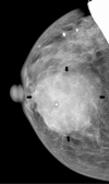
Figure 1A
Figure 1A
75-year-old woman with primary breast lymphoma. Mammogram of the right breast, CC view. Note a large oval isodense mass (arrows) with indistinct margins at 1 o’clock extending from anterior to posterior depth without associated calcifications. A BB marks the site of palpable abnormality.
Figure 1B
Figure 1B
75-year-old woman with primary breast lymphoma. Mammogram of the right breast, ML view. Note a large oval isodense mass (arrows) with indistinct margins at 1 o’clock extending from anterior to posterior depth without associated calcifications. A BB marks the site of palpable abnormality.
Figure 1C
Figure 1C
75-year-old woman with primary breast lymphoma. Mammogram of the right breast, spot magnification, CC view. Note a large oval isodense mass (arrows) with indistinct margins at 1 o’clock extending from anterior to posterior depth without associated calcifications. A BB marks the site of palpable abnormality.
Figure 1D
Figure 1D
75-year-old woman with primary breast lymphoma. Mammogram of the right breast, spot magnification, ML view. Note a large oval isodense mass (arrows) with indistinct margins at 1 o’clock extending from anterior to posterior depth without associated calcifications. A BB marks the site of palpable abnormality.
Figure 2A
Figure 2A
75-year-old woman with primary breast lymphoma. Ultrasound image of the palpable area demonstrates a 44 × 20 × 44 mm heterogeneous, hypoechoic oval mass parallel to the chest wall with circumscribed and microlobulated margins and increased vascularity.
Figure 2B
Figure 2B
75-year-old woman with primary breast lymphoma. Ultrasound image of the palpable area demonstrates a 44 × 20 × 44 mm heterogeneous, hypoechoic oval mass parallel to the chest wall with circumscribed and microlobulated margins and increased vascularity at 1 o'clock 3 cm to the nipple.
Figure 3A
Figure 3A
75-year-old woman with primary breast lymphoma. Ultrasound image of an axillary lymph node reveals loss of the normal reniform appearance with eccentric, bulbous cortical thickening with slitlike configuration of the hilum.
Figure 3B
Figure 3B
75-year-old woman with primary breast lymphoma. Ultrasound image of an axillary lymph node reveals loss of the normal reniform appearance with eccentric, bulbous cortical thickening with slitlike configuration of the hilum.
Figure 3C
Figure 3C
75-year-old woman with primary breast lymphoma. Color Doppler image of the same axillary lymph node demonstrates increased vascularity with increased cortical flow.
Figure 4
Figure 4
Medium power view of the breast biopsy contains a diffuse infiltrate of malignant cells, invading in a sheet-like growth pattern (H&E stain, 200X). The cytologic features of the malignant cells are apparent at higher power. The cells have scant to inapparent cytoplasm with large atypical nuclei containing prominent nucleoli, vesicular chromatin, and irregular nuclear membranes (arrowheads). A normal lymphocyte is bracketed (asterisks) for comparison.

References

    1. D'Orsi C, Bassett L, Berg W. Breast Imaging Reporting and Data System: ACR BI-RADS-Mammography. 4 ed. American College of Radiology; Reston, VA: 2003.
    1. Alvarez S, Anorbe E, Alcorta P, Lopez F, Alonso I, Cortes J. Role of sonography in the diagnosis of axillary lymph node metastases in breast cancer: a systematic review. AJR Am J Roentgenol. 2006;186(5):1342–1348. [PubMed] - PubMed
    1. Stavros AT. Breast Ultrasound. Lippincott Williams & Wilkins; Philadelphia, PA: 2004.
    1. Wong AK, Lopategui J, Clancy S, Kulber D, Bose S. Anaplastic large cell lymphoma associated with a breast implant capsule: a case report and review of the literature. Am J Surg Pathol. 2008;32(8):1265–1268. [PubMed] - PubMed
    1. Espinosa LA, Daniel BL, Jeffrey SS, Nowels KW, Ikeda DM. MRI features of mucosa-associated lymphoid tissue lymphoma in the breast. AJR Am J Roentgenol. 2005;185(1):199–202. [PubMed] - PubMed

Publication types

LinkOut - more resources