

Case Reports
doi: 10.2484/rcr.v5i1.354.
eCollection 2010.
Tightrope walking: A new technique in ankle syndesmosis fixation
- PMID: 27307846
- PMCID: PMC4898177
- DOI: 10.2484/rcr.v5i1.354
Item in Clipboard
Case Reports
Tightrope walking: A new technique in ankle syndesmosis fixation
Radiol Case Rep.
.
Abstract
Over the past few years, several studies have demonstrated favorable clinical outcomes and low complication rates using the ankle Tightrope® syndesmosis fixation system. The traditional surgical procedure of screw fixation for syndesmosis injury is associated with high complication rates of loosening, screw fracture, nonanatomic fixation, and postoperative syndesmotic diastasis. It is expected that the Tightrope® technique will become more common practice given the recent successful reports, so it is important for radiologists to be aware of this novel surgical technique and its imaging appearance.
Keywords: CT, computed tomography.
Figures
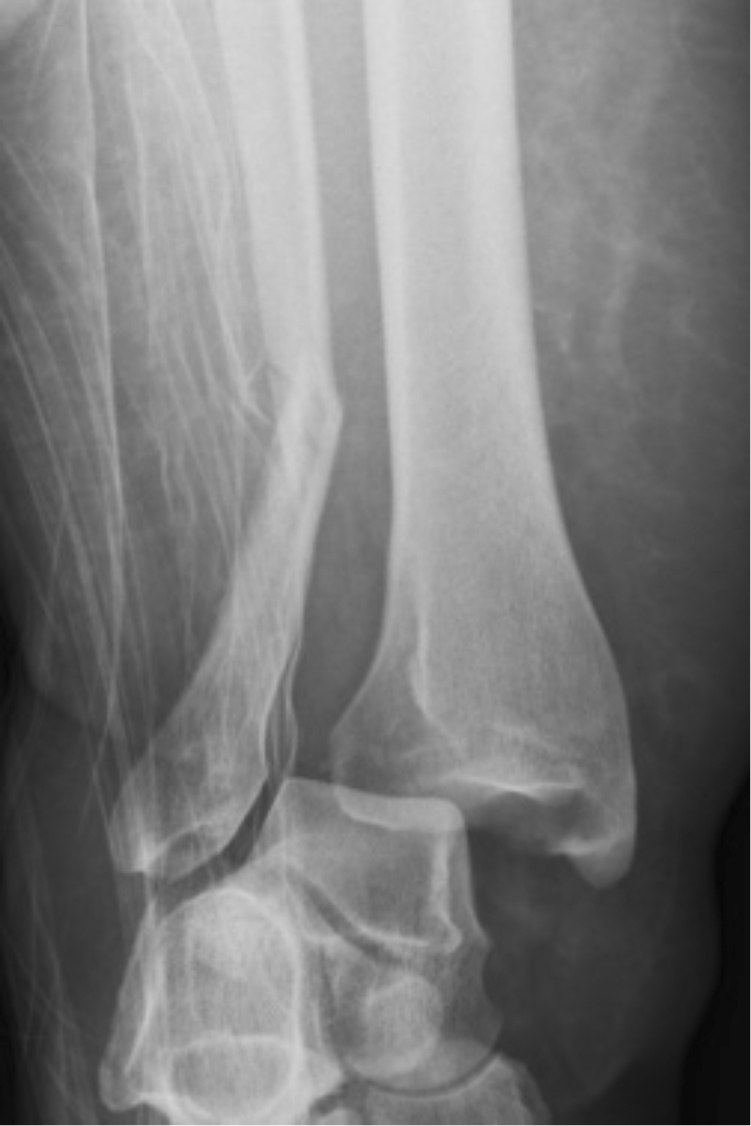
59-year-old woman with right fibular fracture. AP radiograph of the right ankle demonstrates fracture of the distal one-third shaft of the fibula and widening of the distal tibiofibular joint with the tibia medially dislocated. Findings are consistent with a Weber C fracture with syndesmotic disruption.
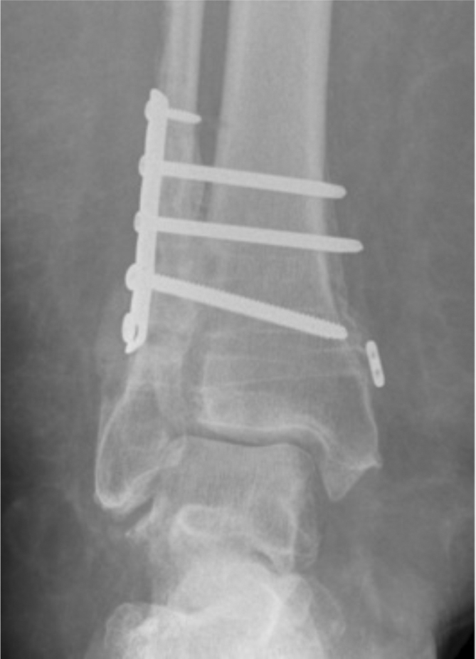
59-year-old woman with right fibular fracture. Plain radiograph of the right ankle shows lateral plate and screw internal fixation of the distal fibula fracture, which has healed. There are also three syndesmotic screws with surrounding lucency, suggestive of loosening. Widening of the distal tibiofibular joint and heterotopic ossification is seen.
59-year-old woman with right fibular fracture. Coronal-plane weight-bearing CT of the right ankle in bone window demonstrates widening of the distal tibiofibular joint.
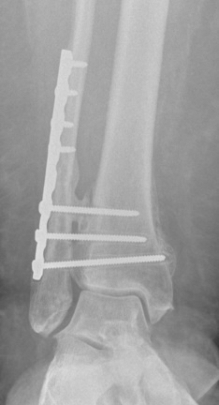
59-year-old woman with right fibular fracture. AP radiograph of the right ankle demonstrates a new lateral fibular plate with one fibular screw and three syndesmotic screws. A radiolucent transverse tunnel is seen parallel to the plafond, consistent with the Tightrope® suture system. An oblong button is present along the medial tibia cortex, and a round button is present against the distal hole of the fibular plate. Widening of the distal tibiofibular joint has been reduced.
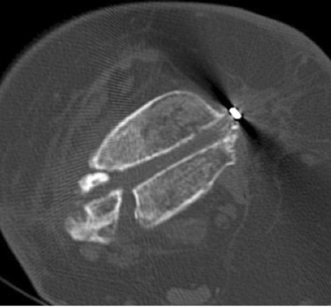
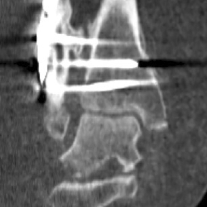
59-year-old woman with right fibular fracture. Axial CT of the right ankle in bone window demonstrates a round metal button along the medial tibial cortex and a low attenuation tunnel extending along the tibiofibular joint.
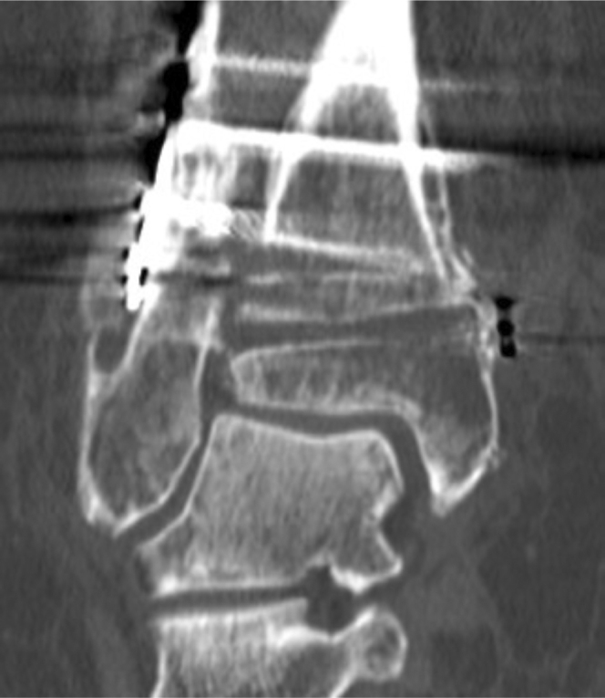
59-year-old woman with right fibular fracture. Coronal CT of the right ankle in bone window demonstrates the new Tightrope® tunnel and normal distal tibiofibular joint space.
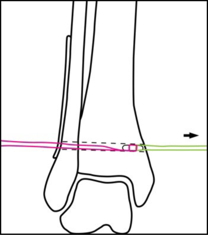
59-year-old woman with right fibular fracture. The oblong button and fiberwire (pink) are passed through the drill hole by the pull-through suture (green).
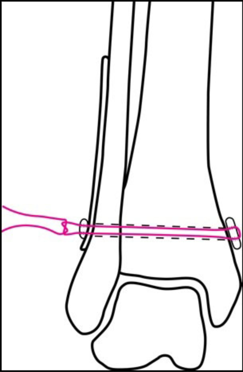
59-year-old woman with right fibular fracture. The oblong button has been flipped perpendicular to the drill hole, and the pull-through suture has been removed. The round lateral button has been threaded over the fiberwire and secured adjacent to the lateral fibular plate with a knot.
References
Publication types
LinkOut - more resources
Full Text Sources
Miscellaneous
