Case Reports
. 2015 Nov 6;5(1):355.
doi: 10.2484/rcr.v5i1.355. eCollection 2010.

Madura foot masquerading as a hemangioma

Case Reports

Madura foot masquerading as a hemangioma

Jonelle M Petscavage et al. Radiol Case Rep. .

Abstract

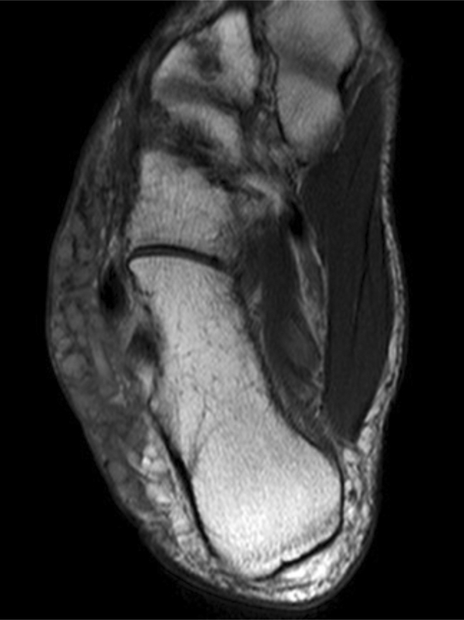
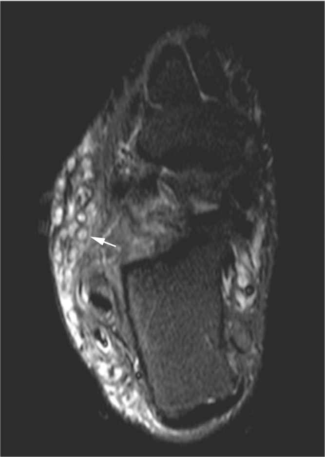
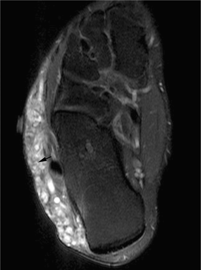
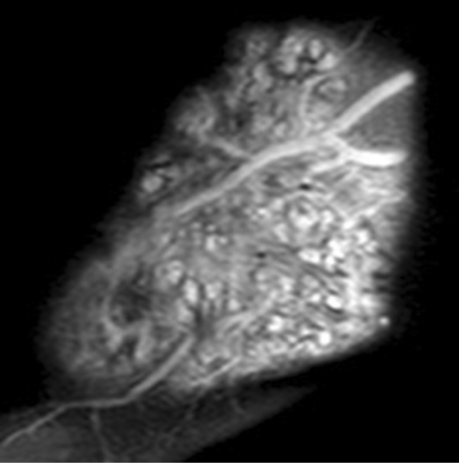
Mycetoma, also known as Madura foot, is a rare soft-tissue granulomatous infection caused by Actinomyces or true fungi. The MRI "dot-in-circle" sign has been described as a characteristic finding of mycetoma. This sign represents spherical T2 bright masses containing central and intervening low-signal-intensity foci. However, other soft-tissue masses can have similar appearances. We present a case of a Madura foot that was erroneously given the imaging diagnosis of soft-tissue hemangioma due to the presence of serpiginous enhancing masses with the "dot-in-circle" sign (believed to be due to phleboliths).

Keywords: CT, computed tomography; MRI, magnetic resonance imaging.


Figures

Figure 1
Figure 1
Axial T1 image of the foot demonstrating round and serpiginous intermediate-signal-intensity masses in the lateral subcutaneous tissues, Some of the masses have internal areas of low T1 signal.
Figure 2
Figure 2
Axial STIR image demonstrates the “dot-in-circle” sign described in mycetoma (arrow).
Figure 3
Figure 3
Axial T1 fat-suppressed image, post contrast, demonstrates enhancement of the mass involving the lateral soft tissues of the foot and “dot-in-circle” sign (arrow).
Figure 4
Figure 4
Sagittal T1 fat-suppressed postgadolinium image at the lateral aspect of the ankle and foot demonstrates a large enhancing mass containing spherical and serpiginous components that contain both central and intervening areas of low signal.

References

    1. Sharif HS. Mycetoma: comparison of MR imaging with CT. Radiology. 1991;178:865–870. [PubMed] - PubMed
    1. Magana M. Mycetoma, some clinical and histopathological features. Turk J Dermatopathol. 1994;3:94.
    1. Magana M. Mycetoma. Int J Dermatol. 1984;23:221–236. [PubMed] - PubMed
    1. McGinnis MR, Fader RC. Mycetoma: a contemporary concept. Infect Dis Clin North Am. 1988;2:939–954. [PubMed] - PubMed
    1. Bendl BJ, Mackey D, Al-Saati F, Sheth Ky, Ofole SN, Baily TM. Mycetoma in Saudi Arabia. J Trop Med Hyg. 1987;90:51–59. [PubMed] - PubMed

Publication types

LinkOut - more resources