

Case Reports
doi: 10.2484/rcr.v5i2.388.
eCollection 2010.
Large omental cyst
- PMID: 27307861
- PMCID: PMC4898220
- DOI: 10.2484/rcr.v5i2.388
Item in Clipboard
Case Reports
Large omental cyst
Radiol Case Rep.
.
Abstract
Omental and mesenteric cysts are rare intra-abdominal masses in the pediatric population. When large, these lesions can mimic large-volume ascites and may not be immediately recognized. This can lead to a delay in appropriate treatment as a result of additional unnecessary tests and procedures. We present a case of a large omental cyst in an 18-month-old male with emphasis on imaging findings that can help diagnose this rare, but important entity.
Keywords: CT, computed tomography.
Figures

18-month-old male with large omental cyst. Image from patient’s initial ultrasound evaluation shows intraperitoneal multilocular fluid collections.
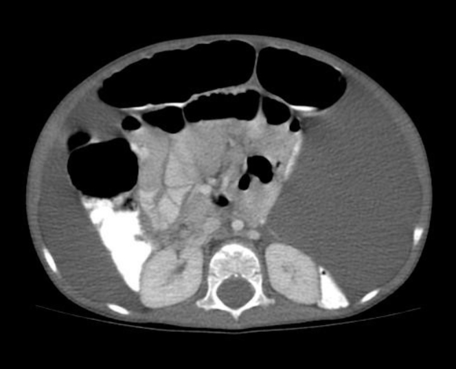
18-month-old male with large omental cyst. Axial CT with IV and oral contrast demonstrates a large amount of low-attenuation intra‐abdominal fluid. The mass effect on the bowel and kidney on the left abdomen suggest a cystic mass lesion. The apparently free fluid also present along the liver persuaded the clinicians to evaluate extensively for a cause for ascites.

18-month-old male with large omental cyst. Coronal CT with IV and oral contrast demonstrates a large amount of low-attenuation intra‐abdominal fluid. The mass effect on the bowel and kidney on the left abdomen suggest a cystic mass lesion. The apparently free fluid also present along the liver persuaded the clinicians to evaluate extensively for a cause for ascites.

18-month-old male with large omental cyst. Axial CT image performed later in the patient’s course demonstrates mass effect in the left abdomen as well as central bowel displacement.

18-month-old male with large omental cyst. Coronal CT image performed later in the patient’s course demonstrates mass effect in the left abdomen as well as central bowel displacement.

18-month-old male with large omental cyst. Ultrasound images again show a multilocular fluid collection in the abdomen.

18-month-old male with large omental cyst. Ultrasound images again show a multilocular fluid collection in the abdomen with nondependent bowel loops in the right lower quadrant.
References
Publication types
LinkOut - more resources
Full Text Sources
