Case Reports
. 2015 Nov 6;5(3):394.
doi: 10.2484/rcr.v5i3.394. eCollection 2010.

Pseudolesion in segment IV A of the liver from vein of Sappey secondary to SVC obstruction

Case Reports

Pseudolesion in segment IV A of the liver from vein of Sappey secondary to SVC obstruction

Dharshan Vummidi et al. Radiol Case Rep. .

Abstract

Pseudolesions in the liver are caused by unusual/altered hemodynamics of the liver and can be confused with a true hepatic mass. In superior vena cava (SVC) obstruction. there is recruitment of the cavo-mammary-phrenic-hepatic-capsule-portal pathway. and the venous blood follows the internal mammary vein, the inferior phrenic vein, the hepatic capsule veins, and the intrahepatic portal system. causing a hypervascular pseudolesion in segment IV A of the liver. Recognizing the classic appearances of this hypervascular pseudolesion from the vein of Sappey in a CT study of the abdomen has prognostic implications in directing further evaluation of the chest for SVC obstruction. We present a case of a 54-year-old HIV-positive male smoker in whom identification of the hypervascular pseudolesion from the vein of Sappey on the abdominal CT led to the diagnosis of SVC syndrome.

Keywords: CT, computed tomography; SVC, superior vena cava.


Figures

Figure 1A, B
Figure 1A, B
54-year-old HIV-positive man with hypervascular pseudolesion in segment IV A and vein of Sappey from SVC obstruction. Axial CT images demonstrate hypervascular pseudolesion (arrow) in the segment IV A with enhancing vein of Sappey (arrowhead). Also note collateral recruitment of azygos and hemiazygos veins in addition to cutaneous collaterals. There are bilateral, right-greater-than-left pleural effusions..
Figure 1A, B
Figure 1A, B
54-year-old HIV-positive man with hypervascular pseudolesion in segment IV A and vein of Sappey from SVC obstruction. Axial CT images demonstrate hypervascular pseudolesion (arrow) in the segment IV A with enhancing vein of Sappey (arrowhead). Also note collateral recruitment of azygos and hemiazygos veins in addition to cutaneous collaterals. There are bilateral, right-greater-than-left pleural effusions..
Figure 1C
Figure 1C
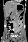
54-year-old HIV-positive man with hypervascular pseudolesion in segment IV A and vein of Sappey from SVC obstruction. Coronal CT image demonstrates hypervascular pseudolesion (arrow) in the segment IV A. The vein of Sappey was best visualized on the axial images.
Figure 2A, B
Figure 2A, B
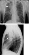
54-year-old HIV-positive man with hypervascular pseudolesion in segment IV A and vein of Sappey from SVC obstruction. Frontal (A) and lateral (B) chest radiographs demonstrate right upper lobe mass (arrow) with enlarged right paratracheal lymph nodes (arrowhead) and mass effect on the SVC.
Figure 3A, B
Figure 3A, B
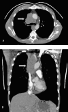
54-year-old HIV-positive man with hypervascular pseudolesion in segment IV A and vein of Sappey from SVC obstruction. Axial (A) and coronal (B) CT images through the chest demonstrate a mediastinal mass obstructing the superior vena cava (arrow) with extensive prominent collaterals in the left chest wall and scapular region.
Figure 4
Figure 4
54-year-old HIV-positive man with hypervascular pseudolesion in segment IV A and vein of Sappey from SVC obstruction. Hematoxylin & Eosin stain histopathology image demonstrates numerous. small. highly atypical cells with high nuclear-cytoplasmic volume ratios consistent with small-cell carcinoma of the lung.

References

    1. Salsali M, Cliffton EE. Superior vena caval obstruction in carcinoma of lung. N Y State J Med. 1969;69(22):2875–2880. [PubMed] - PubMed
    1. Escalante CP. Causes and management of superior vena cava syndrome. Oncology:Williston Park. 1993;7:61–68. - PubMed
    1. Nieto AF, Doty DB. Superior vena cava obstruction; clinical syndrome, etiology and treatment. Curr Probl Cancer. 1986;10(9):441–484. [PubMed] - PubMed
    1. Lee JW, Kim S, Kwack SW. Hepatic Capsular and subcapsular pathologic conditions: Demonstration with CT and MR imaging. RadioGraphics. 2008 Sep-Oct;28(5):1307–1323. [PubMed] - PubMed
    1. Matsui O, Takahashi S, Kadoya M. Pseudolesion in segment IV of the liver at CT during arterial portography: correlation with aberrant gastric venous drainage. Radiology. 1994 Oct;193(1):31–35. [PubMed] - PubMed

Publication types

LinkOut - more resources