

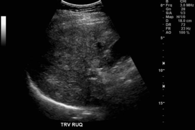
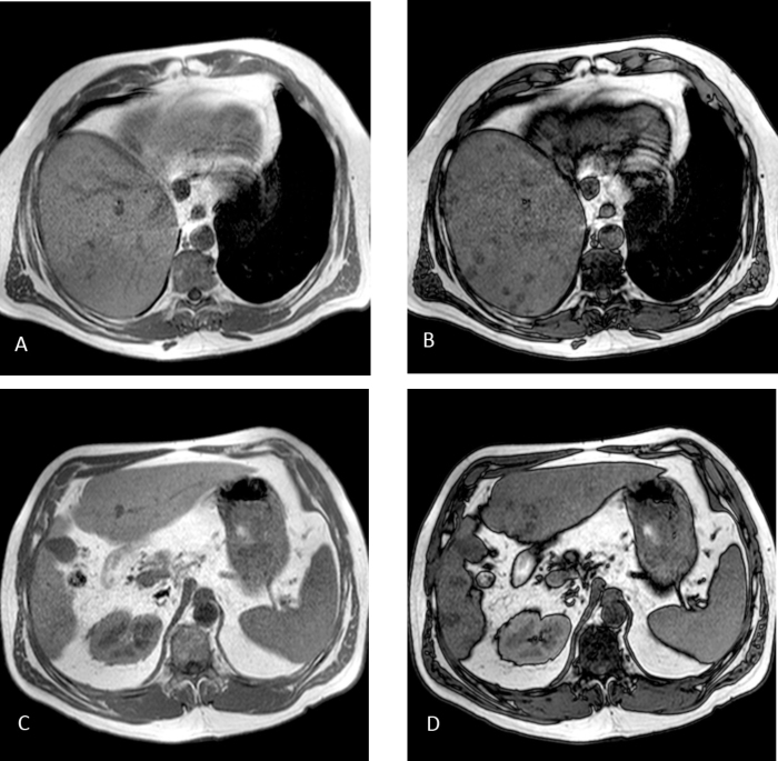
Multiple echogenic liver masses from multifocal nodular steatosis in a 55-year-old male with chronic hepatitis C
- PMID: 27307868
- PMCID: PMC4898294
- DOI: 10.2484/rcr.v5i3.399
Multiple echogenic liver masses from multifocal nodular steatosis in a 55-year-old male with chronic hepatitis C
Abstract
Hepatic steatosis is often seen in patients with hepatitis on screening ultrasound as generalized increased liver echogenecity. However, its nodular form can present as multiple echogenic masses, which can mimic hepatocellular carcinoma or metastasis by ultrasound and computed tomography. Small hepatocellular carcinomas are often hyperechoic and have a trend towards lower alpha-fetoprotein levels. Magnetic resonance imaging can accurately identify microscopic fat within the lesions and demonstrate lack of associated enhancing soft tissue. If this entity is not appropriately characterized using magnetic resonance imaging, it can lead to additional imaging workup and unnecessary biopsy.
Keywords: AFP, alpha-fetoprotein; AML, angiomyolipoma; CT, computed tomography; FNH, focal nodular hyperplasia; HCC, hepatocellular carcinoma; MRI, magnetic resonance imaging; US, ultrasound.
Figures


References
Publication types
LinkOut - more resources
Full Text Sources
