

Case Reports
doi: 10.2484/rcr.v6i3.486.
eCollection 2011.
Hydatid cyst: Anywhere, everywhere
- PMID: 27307908
- PMCID: PMC4900053
- DOI: 10.2484/rcr.v6i3.486
Item in Clipboard
Case Reports
Hydatid cyst: Anywhere, everywhere
Radiol Case Rep.
.
Abstract
Hydatid cyst is a parasitic infection that primarily affects the liver but which can be found anywhere in the body. This case involves spontaneous dissemination of hydatid cyst disease, a rare occurrence in the absence of any intervention or trauma.
Keywords: CT, computed tomography; HD, hydatid disease.
Figures

35-year-old male with complaints of abdominal distention. Ultrasound of abdomen using 5 MHz curvilinear probe shows in the mesentry and omentum.
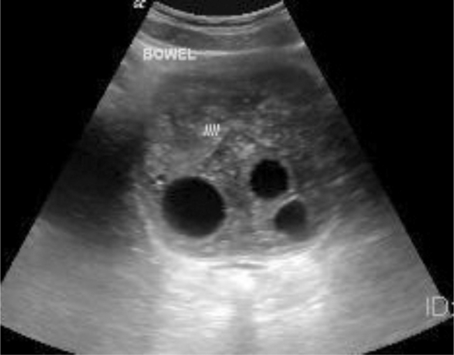
35-year-old male with complaints of abdominal distention. Ultrasound of abdomen using 5 MHz curvilnear probe shows multiple cystic lesions in the mesentry and omentum.
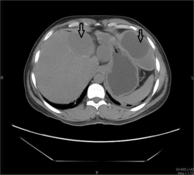
35-year-old male with complaints of abdominal distention. Axial noncontrast-enhanced CT of the abdomen shows multiple cystic lesions in the omentum and liver (kvp :120 mas: 100, 5mm thickness).

35-year-old male with complaints of abdominal distention. Axial noncontrast-enhanced CT of the abdomen shows multiple cystic lesions in the pelvis in the mesentery and near the bladder (Kvp:120 mas:100 5mm thickness).

35-year-old male with complaints of abdominal distention. Axial noncontrast-enhanced CT of the thorax in mediastinal (A) and lung window (B) shows a large cyst in the left posterobasal segment of the left lobe with broncheiectatic changes in the right lower lobe. Another cyst seen adjacent to the spleen. (Kvp: 120 mas:100 5 mm thickness).
References
-
- Pumarola A, Rodriguez-Torres A, García-Rodriguez JA, Piédrola-Angulo G. Microbiología y parasitología médica. 2nd ed. Salvat; Barcelona, Spain: 1990.
-
- King CH. Cestodes (tapeworms) In: Mandell GL, Bennett JE, Dolin R, editors. Principles and practice of infectious diseases. 4th Ed. Churchill Livingstone; New York, NY: 1995. pp. 2544–2553.
Publication types
LinkOut - more resources
Full Text Sources
