Postnatal Evaluation and Outcome of Prenatal Hydronephrosis
- PMID: 27307966
- PMCID: PMC4906562
- DOI: 10.5812/ijp.3667
Postnatal Evaluation and Outcome of Prenatal Hydronephrosis
Abstract
Background: Prenatal hydronephrosis (PNH) is dilation in urinary collecting system and is the most frequent neonatal urinary tract abnormality with an incidence of 1% to 5% of all pregnancies. PNH is defined as anteroposterior diameter (APD) of renal pelvis ≥ 4 mm at gestational age (GA) of < 33 weeks and APD ≥ 7 mm at GA of ≥ 33 weeks to 2 months after birth. All patients need to be evaluated after birth by postnatal renal ultrasonography (US). In the vast majority of cases, watchful waiting is the only thing to do; others need medical or surgical therapy.
Objectives: There is a direct relationship between APD of renal pelvis and outcome of PNH. Therefore we were to find the best cutoff point APD of renal pelvis which leads to surgical outcome.
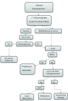
Patients and methods: In this retrospective cohort study we followed 200 patients 1 to 60 days old with diagnosis of PNH based on before or after birth ultrasonography; as a prenatal or postnatal detected, respectively. These patients were referred to the nephrology clinic in Zahedan Iran during 2011 to 2013. The first step of investigation was a postnatal renal US, by the same expert radiologist and classifying the patients into 3 groups; normal, mild/moderate and severe. The second step was to perform voiding cystourethrogram (VCUG) for mild/moderate to severe cases at 4 - 6 weeks of life. Tc-diethylene triamine-pentaacetic acid (DTPA) was the last step and for those with normal VCUG who did not show improvement in follow-up examination, US to evaluate obstruction and renal function. Finally all patients with mild/moderate to severe PNH received conservative therapy and surgery was preserved only for progressive cases, obstruction or renal function ≤35%. All patients' data and radiologic information was recorded in separate data forms, and then analyzed by SPSS (version 22).
Results: 200 screened PNH patients with male to female ratio 3.5:1 underwent first postnatal control US, of whom 65% had normal, 18% mild/moderate and 17% severe hydronephrosis. 167 patients had VCUG of whom 20.82% with VUR. 112 patients performed DTPA with following results: 50 patients had obstruction and 62 patients showed no obstructive finding. Finally 54% of 200 patients recovered by conservative therapy, 12.5% by surgery and remaining improved without any surgical intervention.
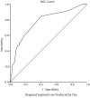
Conclusions: The best cutoff point of anteroposterior renal pelvis diameter that led to surgery was 15 mm, with sensitivity 88% and specificity 74%.
Keywords: Prenatal Hydronephrosis; Renal Pelvis; Ultrasonography.
Figures
References
-
- Sharifian M, Esfandiar N, Mohkam M, Dalirani R, Baban Taher E, Akhlaghi A. Diagnostic accuracy of renal pelvic dilatation in determining outcome of congenital hydronephrosis. Iran J Kidney Dis. 2014;8(1):26–30. - PubMed
-
- Molina CA, Facincani I, Muglia VF, Araujo WM, Cassini MF, Tucci Jr S. Postnatal evaluation of intrauterine hydronephrosis due to ureteropelvic junction obstruction. Acta Cir Bras. 2013;28 Suppl 1:33–6. - PubMed
LinkOut - more resources
Full Text Sources
Other Literature Sources


