Review
. 2014 Oct 30;2(1):e968020.
doi: 10.4161/23723548.2014.968020. eCollection 2015 Jan-Mar.

Role of the double-strand break repair pathway in the maintenance of genomic stability

Affiliations
Review

Role of the double-strand break repair pathway in the maintenance of genomic stability

Tangui Le Guen et al. Mol Cell Oncol. .

Abstract

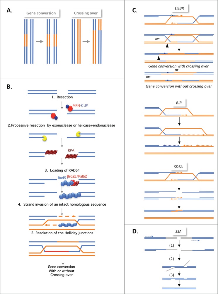
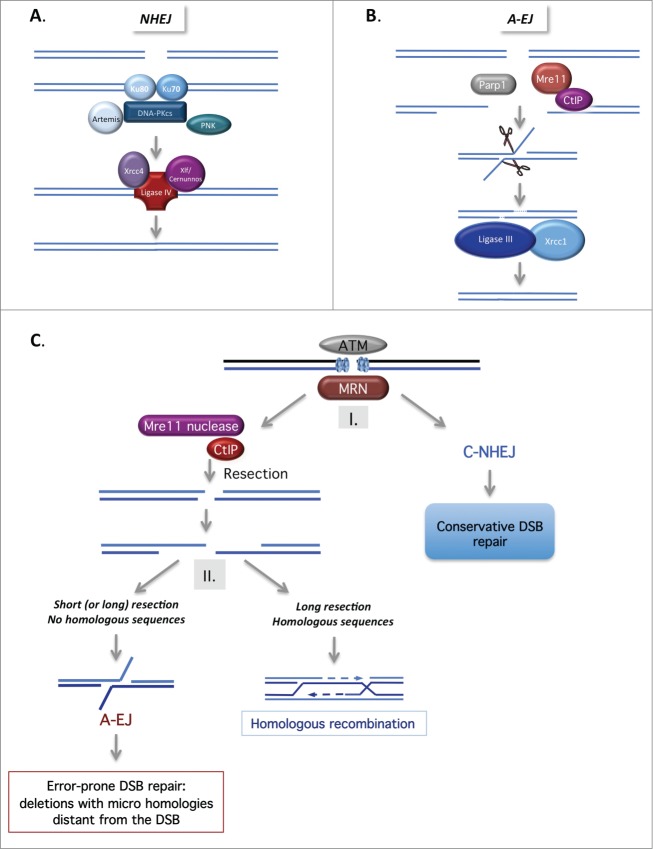
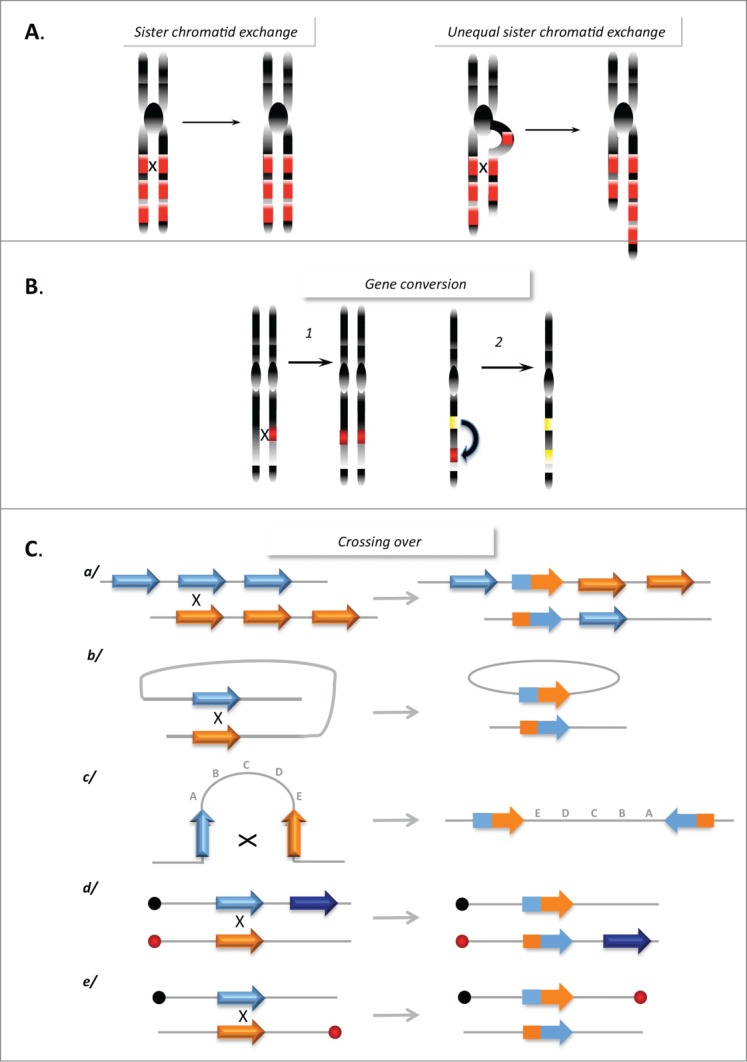
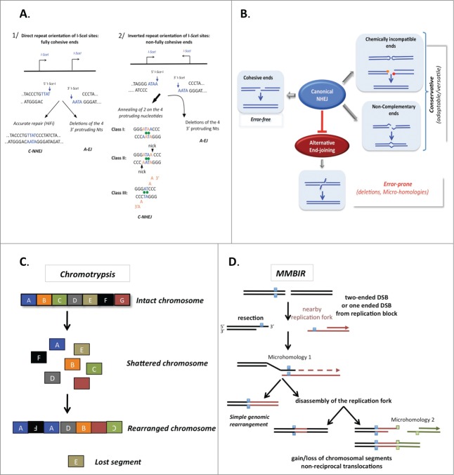
DNA double-strand breaks (DSBs) are highly lethal lesions that jeopardize genome integrity. However, DSBs are also used to generate diversity during the physiological processes of meiosis or establishment of the immune repertoire. Therefore, DSB repair must be tightly controlled. Two main strategies are used to repair DSBs: homologous recombination (HR) and non-homologous end joining (NHEJ). HR is generally considered to be error-free, whereas NHEJ is considered to be error-prone. However, recent data challenge these assertions. Here, we present the molecular mechanisms involved in HR and NHEJ and the recently described alternative end-joining mechanism, which is exclusively mutagenic. Whereas NHEJ is not intrinsically error-prone but adaptable, HR has the intrinsic ability to modify the DNA sequence. Importantly, in both cases the initial structure of the DNA impacts the outcome. Finally, the consequences and applications of these repair mechanisms are discussed. Both HR and NHEJ are double-edged swords, essential for maintenance of genome stability and diversity but also able to generate genome instability.

Keywords: DNA repair; double-strand break repair; genome instability; genome rearrangements; homologous recombination; ionizing radiation; mutagenesis; non-homologous end joining; telomeres.


Figures

Figure 1
Figure 1
(See previous page). Mechanisms of homologous recombination. (A) The products of HR. Gene conversion (GC; left panel), results from the non-reciprocal exchange of a DNA sequence (in orange). Crossing over (CO; right panel), results from the reciprocal exchange of DNA sequences (orange and blue). Both GC and CO are outcomes of the HR events described below. (B) The double strand break repair model (DSBR). HR is initiated by 5′ to 3′ single-stranded resection of double-stranded DNA ends through the action of different protein complexes, MRN/CtIP, Exo1, and BLM/TopoIII/RMI, acting in 2 steps. BRCA1, in association with CtIP, favors resection initiation through the removal of 53BP1. This resection creates a 3′ single-stranded DNA (ssDNA) that is coated with replication protein A (RPA). A complex including Palb2 and BRCA2, which are both breast tumor suppressors, then replaces RPA with Rad51. The Rad51 nucleoprotein filament invades the intact homologous duplex DNA, priming DNA synthesis, and the intact DNA molecule is copied, creating a D-Loop (displacement loop). This process tolerates limited polymorphisms, thus creating heteroduplex intermediates bearing mismatches (blue circle in corresponding left panel in Fig. 1C). This step generates 2 cruciform intermediates also known as Holliday junctions. (C) Different HR mechanisms. In the DSBR model (upper panel), the heteroduplex molecules are represented by the blue circles. Strand invasion and DNA synthesis can lead to GCs. Depending on how the Holliday junctions are resolved, the GC will be associated with CO (black arrow) or not (gray arrow). In synthesis-dependent strand annealing (SDSA, middle panel), the initiation is similar but the invading strand dehybridizes and reanneals at the other end of the broken molecule and no Holliday junction is generated. In break-induced replication (BIR, lower panel), DNA synthesis occurs over longer distances, even reaching the end of the chromosome. Here, there is neither resolution of the intermediates nor crossing over. (C) Single-strand annealing (SSA). An extended single-strand resection (1) reveals 2 complementary ssDNA strands; hybridization of the 2 complementary strands (2) generates an intermediate; resolution (3) and gap filling complete the repair. This process can occur between 2 homologous sequences in tandem in the same orientation (dotted arrows) and results in deletion of the intergenic sequences.
Figure 2
Figure 2
(See previous page). Mechanisms of non-homologous end joining. (A). Canonical NHEJ (C-NHEJ). C-NHEJ is initiated by binding of the Ku80-Ku70 heterodimer to the DSB, which recruits the DNA-PK catalytic subunit, DNA-PKcs. Several proteins, including Artemis, the polynucleotide kinase (PNK) and members of the polymerase X family, process the DNA ends to make them competent for the subsequent ligation steps. Finally, ligase IV, in association with Xrcc4 and Cernunos/Xlf, seals the double-strand ends. (B) Alternative end-joining (A-EJ). In the absence of Ku70/Ku80, the DNA ends are resected in a reaction favored by the nuclease activity of Mre11 and CtIP. Note that Parp1 is involved in A-EJ initiation. The resulting ssDNA reveals complementary microhomologies (2–4 nt or more) that can be annealed; gap filling completes the end joining. Finally, Xrcc1 and ligase III complete the A-EJ process. Notably, A-EJ is always associated with deletions at the junctions and frequently, but not systematically, involves microhomologies that are distant from the DSB. (C) The 2-step DSB repair pathway choice model. After signaling of the DSB by ATM and MRN, (I) binding of Ku80/Ku70 protects the DSB ends from resection, routing DSB repair toward the conservative C-NHEJ pathway. (II) The nuclease activity of Mre11 and CtIP favor ssDNA resection, which can then initiate the HR or A-EJ pathway. A short ssDNA resection is sufficient for A-EJ but not for HR. A-EJ is an exclusively non-conservative mutagenic process.
Figure 3.
Figure 3.
Genome instability promoted by HR. (A) Sister chromatid exchanges. Left panel: Equal SCE between repeat sequences (red boxes) does not affect genome stability. Right panel: Unequal SCEs, leading to amplification and loss of genetic material. (B) Gene conversion between 2 heteroalleles (left panel) leading to a loss of heterozygosity, or between a pseudogene (yellow box) and a gene (red box) leading to inactivation of the gene. (C) Rearrangements resulting from CO between repeat sequences: (a) Between homologous sequences on 2 chromosomes, leading to amplification and deletion; (b) Intramolecular CO between direct repeats, resulting in the excision of the intervening sequence; (c) Intramolecular CO between 2 inverted repeats, resulting in inversion of the internal fragment; (d and e) Interchromosomal CO generates a translocation (d) or a dicentric and an acentric chromosome (e) depending on the position relative to their respective centromeres (black and red circles).
Figure 4.
Figure 4.
The accuracy of end joining. (A) The fidelity of C-NHEJ on fully (left panel) or incompletely (right panel) complementary ends. Here, examples are given with ends generated by the meganuclease I-SceI. The cleavage sites are not palindromic therefore 2 I-SceI cleavage sites in the same orientations yield fully complementary ends (left panel), whereas 2 I-SceI cleavage sites in inverted orientation yield incompletely complementary overhangs (right panel). A-EJ leads to deletions with both types of DNA ends. C-NHEJ is mainly error-free on fully complementary ends and uses 3 classes of imperfect annealing (3 out of the 4 3′ protruding nucleotides generated by I-SceI cleavage) with non-fully complementary ends. Thus, C-NHEJ is conservative but adaptable for incompletely complementary ends. (B) The actual accuracy of end joining. A-EJ is highly mutagenic in all situations but it is blocked by C-NHEJ, which can act on imperfectly complementary ends. In situations producing non-ligatable ends, such as hairpins in V(D)J recombination or IR-induced multiple damages at DSBs, a preliminary processing step is required prior to end joining. Note that in these cases, diversity or mutagenesis is generated by the processing step rather than the end-joining machinery. (C) Chromothripsis. The religation of shattered chromosomes in small pieces (colored squares) leads to a combination of rearranged chromosomes in addition to amplification and loss of the small pieces. (D) A model for chromothripsis arising through MMBIR (microhomology-mediated break induced replication). Resection of the DNA double-strand end generates a 3′ overhang, which can anneal another DNA molecule (in red) through the use of microhomologies (blue squares), thus priming DNA synthesis (dotted arrow). This mechanism can lead to more complex rearrangements if it is coupled to multiple cycles of disassembly and template switches of the replication forks (right panel), still using microhomologies.

References

    1. Haber JE. Genome stability. DNA Repair Recombination. Talor and Francis Group: New York; 2014.
    1. Magdalou I, Lopez BS, Pasero P, Lambert SA. The causes of replication stress and their consequences on genome stability and cell fate. Semin Cell Dev Biol 2014; 30C:154-64; http://dx.doi.org/10.1016/j.semcdb.2014.04.035 - DOI - PubMed
    1. Betermier M, Bertrand P, Lopez BS. Is non-homologous end-joining really an inherently error-prone process? PLoS Genet 2014; 10:e1004086; PMID: 24453986; http://dx.doi.org/10.1371/journal.pgen.1004086 - DOI - PMC - PubMed
    1. Rass E, Grabarz A, Plo I, Gautier J, Bertrand P, Lopez BS. Role of Mre11 in chromosomal nonhomologous end joining in mammalian cells. Nat Struct Mol Biol 2009; 16:819-24; PMID: 19633668; http://dx.doi.org/10.1038/nsmb.1641 - DOI - PubMed
    1. Pierce AJ, Hu P, Han M, Ellis N, Jasin M. Ku DNA end-binding protein modulates homologous repair of double-strand breaks in mammalian cells. Genes Dev 2001; 15:3237-42; PMID: 11751629; http://dx.doi.org/10.1101/gad.946401 - DOI - PMC - PubMed

LinkOut - more resources