. 2016 Jun 16;11(6):e0156805.
doi: 10.1371/journal.pone.0156805. eCollection 2016.

Distinct BOLD fMRI Responses of Capsaicin-Induced Thermal Sensation Reveal Pain-Related Brain Activation in Nonhuman Primates

Affiliations

Distinct BOLD fMRI Responses of Capsaicin-Induced Thermal Sensation Reveal Pain-Related Brain Activation in Nonhuman Primates

Abu Bakar Ali Asad et al. PLoS One. .

Abstract

Background: Approximately 20% of the adult population suffer from chronic pain that is not adequately treated by current therapies, highlighting a great need for improved treatment options. To develop effective analgesics, experimental human and animal models of pain are critical. Topically/intra-dermally applied capsaicin induces hyperalgesia and allodynia to thermal and tactile stimuli that mimics chronic pain and is a useful translation from preclinical research to clinical investigation. Many behavioral and self-report studies of pain have exploited the use of the capsaicin pain model, but objective biomarker correlates of the capsaicin augmented nociceptive response in nonhuman primates remains to be explored.

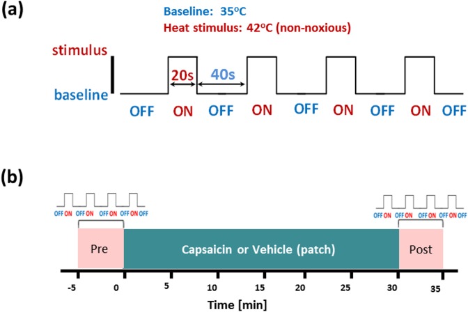
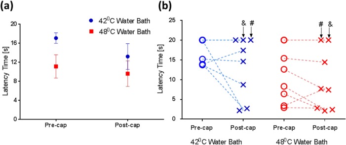
Methodology: Here we establish an aversive capsaicin-induced fMRI model using non-noxious heat stimuli in Cynomolgus monkeys (n = 8). BOLD fMRI data were collected during thermal challenge (ON:20 s/42°C; OFF:40 s/35°C, 4-cycle) at baseline and 30 min post-capsaicin (0.1 mg, topical, forearm) application. Tail withdrawal behavioral studies were also conducted in the same animals using 42°C or 48°C water bath pre- and post- capsaicin application (0.1 mg, subcutaneous, tail).

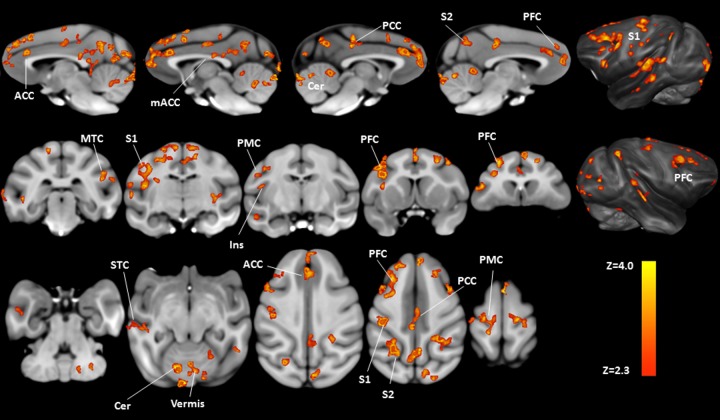
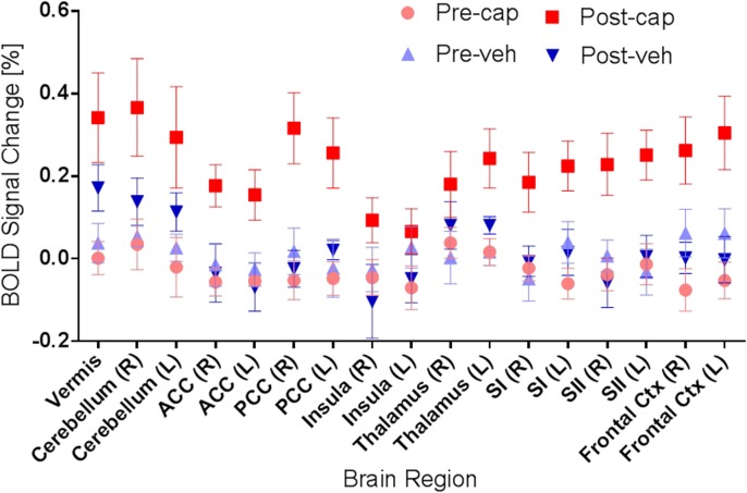
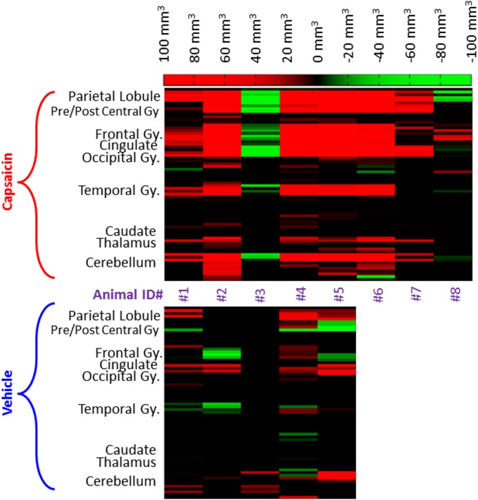
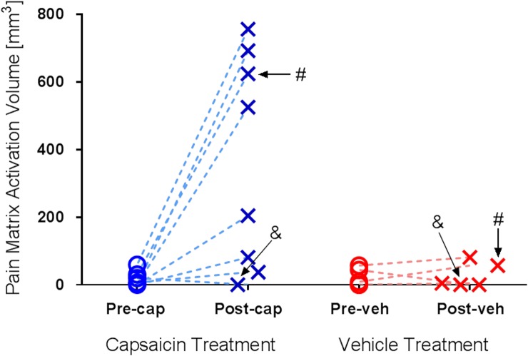
Principal findings: Group comparisons between pre- and post-capsaicin application revealed significant BOLD signal increases in brain regions associated with the 'pain matrix', including somatosensory, frontal, and cingulate cortices, as well as the cerebellum (paired t-test, p<0.02, n = 8), while no significant change was found after the vehicle application. The tail withdrawal behavioral study demonstrated a significant main effect of temperature and a trend towards capsaicin induced reduction of latency at both temperatures.

Conclusions: These findings provide insights into the specific brain regions involved with aversive, 'pain-like', responses in a nonhuman primate model. Future studies may employ both behavioral and fMRI measures as translational biomarkers to gain deeper understanding of pain processing and evaluate the preclinical efficacy of novel analgesics.


Conflict of interest statement

Competing Interests: Merck & Co. provided all funds for this work. Some authors are current or former employees of Merck & Co. and may own stock or hold stock options in Merck & Co. Co-author SWGD is an employee of National University of Singapore and EM was an employee of Maccine Pte Ltd. This does not alter the authors' adherence to all the PLOS ONE policies on sharing data and materials.

Figures

Fig 1
Fig 1. Capsaicin pain fMRI experimental protocol with heat challenge.
(a) The heat stimulation paradigm used in the imaging study. The baseline (OFF) temperature was 35°C (40 s), while heat stimulus (ON) at 42°C (20 s) were exerted using a thermode placed at the left forearm of the animal. Four cycles were repeated for each individual trial. (b) The imaging protocol used in the capsaicin-heat BOLD fMRI experiment. Imaging data were acquired during the baseline (‘Pre’) and post -capsaicin or -vehicle patch application (‘Post’). The capsaicin solution was delivered via a patch (0.1 mg of capsaicin in a dosing volume of 0.1 mL) attached at the location where the thermode was placed.
Fig 2
Fig 2. The effect of temperature and capsaicin on tail withdrawal latency.
(a) Our data show that compared to the baseline (‘Pre’), decreases in latency time (mean ± SEM, n = 8) were found after capsaicin application (‘Post’) at both temperatures and the effect of temperature was significant (F1,7 = 7.89, p = 0.026); however, there was no effect of capsaicin (F1,7 = 2.69, p = 0.145). (b) Tail withdrawal latency time measured from individual animals highlight the inter-subject variability, with two ‘non-responders’ (labelled by ‘#’ and ‘&’) identified (no response to the thermal challenge at both temperatures).
Fig 3
Fig 3. Group comparisons of brain activation patterns showing the effect of capsaicin-induced hypersensitization on heat fMRI signals.
After the topical application of capsaicin, significant increases in BOLD response to 42°C challenge can be found in several pain-related brain regions (pre- versus post-capsaicin application, paired t-test, p<0.02, n = 8). Labeled brain areas in neurological orientation are: ACC = anterior cingulate cortex; mACC = mid anterior cingulate cortex; PCC = posterior cingulate cortex; S2 = secondary somatosensory cortex; PFC = prefrontal cortex; S1 = primary somatosensory cortex; MTC = mid temporal cortex; Ins = insula; PMC = primary motor cortex; STC = superior temporal cortex; Cer = cerebellum.
Fig 4
Fig 4. Regional BOLD signal changes induced by 42°C heat challenge at pre- and post-capsaicin application.
Region-of-interest (ROI) data analyses revealed that significant increases in BOLD signal change (mean ± SEM) was found after the capsaicin (cap) application, but not vehicle (veh) treatment. Specific brain regions associated with pain perception are highlighted.
Fig 5
Fig 5. Inter-subject difference in activated brain volume potentiated by the application of capsaicin or vehicle.
Differences in activated brain volume measured from individual animals, elucidating the inter-subject variability of the capsaicin effect, where red represents an increase in activated brain volume after the capsaicin application and green represents the opposite. As shown, increases in activated brain volume after the capsaicin application can be found in most animals (6 out of 8) whilst the vehicle application did not elicit robust alterations for most animals.
Fig 6
Fig 6. Activated brain volume within the ‘pain matrix’ observed in individual animals.
42°C heat challenge elicited minimal activation within the ‘pain matrix’ in both pre and post -capsaicin (Pre-cap and Post-cap) or -vehicle (Pre-veh and Post-veh). Variation in activated brain volumes under post-capsaicin heat challenge was observed: 4 out of 8 animals showed large increases; another 2 animals showed modest increases, and the remaining 2 animals failed to response to capsaicin challenge. Interestingly, two non-responders identified in the tail withdrawal behavioral assay (‘#’ and ‘&’, see Fig 2) showed differential responses in the imaging study.

References

    1. Breivik H, Collett B, Ventafridda V, Cohen R, Gallacher D. Survey of chronic pain in Europe: prevalence, impact on daily life, and treatment. Eur J Pain. 2006. May;10(4):287–333. - PubMed
    1. Kissin I. The development of new analgesics over the past 50 years: a lack of real breakthrough drugs. Anesth Analg. 2010. March 1;110(3):780–9. 10.1213/ANE.0b013e3181cde882 - DOI - PubMed
    1. Mogil JS. Animal models of pain: progress and challenges. Nat Rev Neurosci. 2009. April;10(4):283–94. 10.1038/nrn2606 - DOI - PubMed
    1. Sandkuhler J. Models and mechanisms of hyperalgesia and allodynia. Physiol Rev. 2009. April;89(2):707–58. 10.1152/physrev.00025.2008 - DOI - PubMed
    1. Chizh BA, Priestley T, Rowbotham M, Schaffler K. Predicting therapeutic efficacy—experimental pain in human subjects. Brain Res Rev. 2009. April;60(1):243–54. 10.1016/j.brainresrev.2008.12.016 - DOI - PubMed

MeSH terms