. 2016 Jun 23;534(7608):570-4.
doi: 10.1038/nature18002. Epub 2016 Jun 15.

Proteome-wide covalent ligand discovery in native biological systems

Affiliations

Proteome-wide covalent ligand discovery in native biological systems

Keriann M Backus et al. Nature. .

Abstract

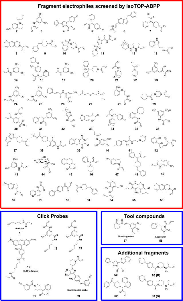
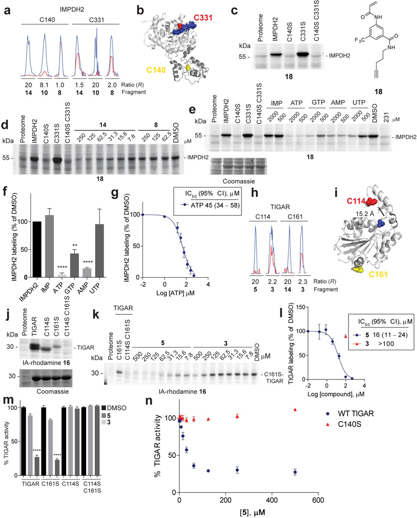
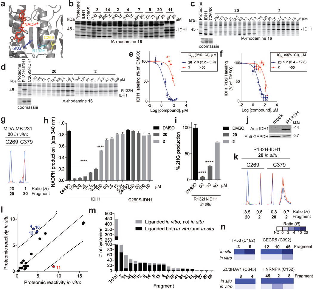
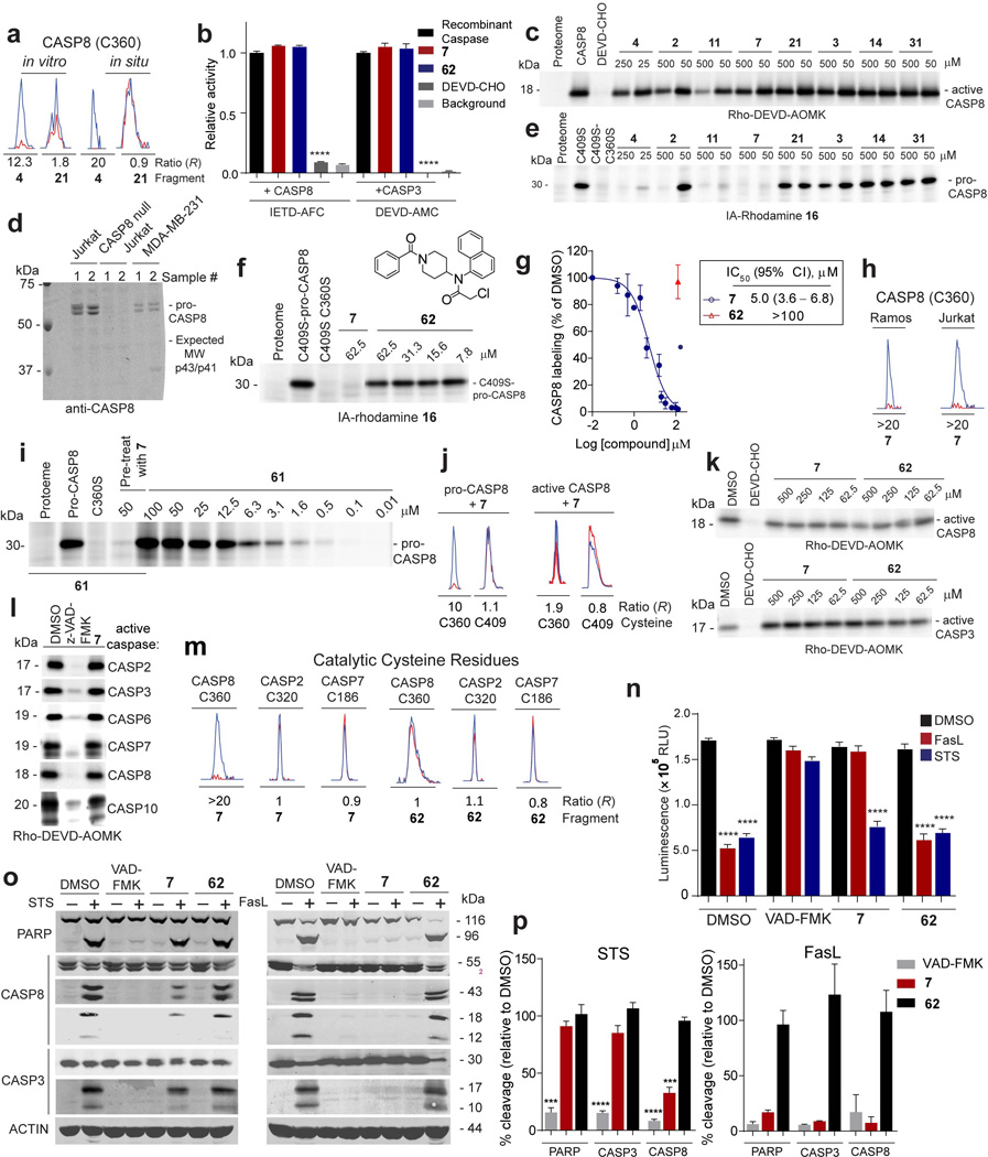
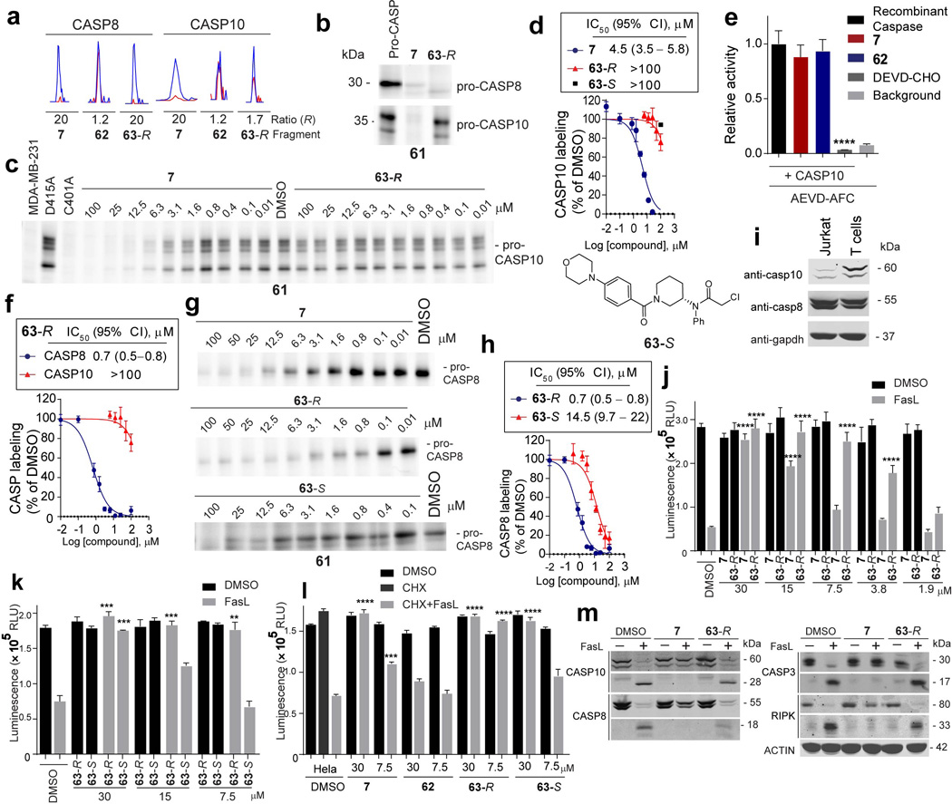
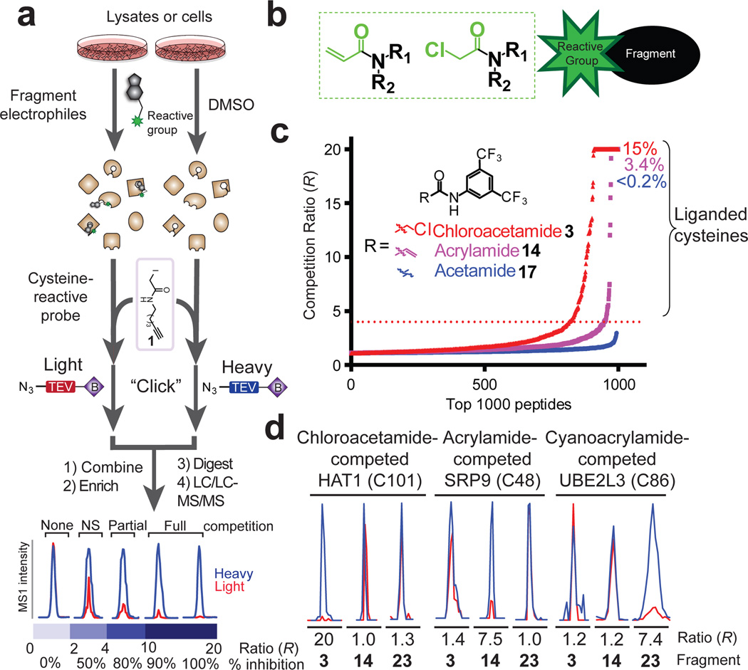
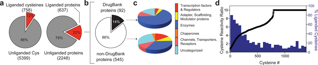
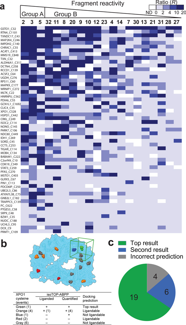
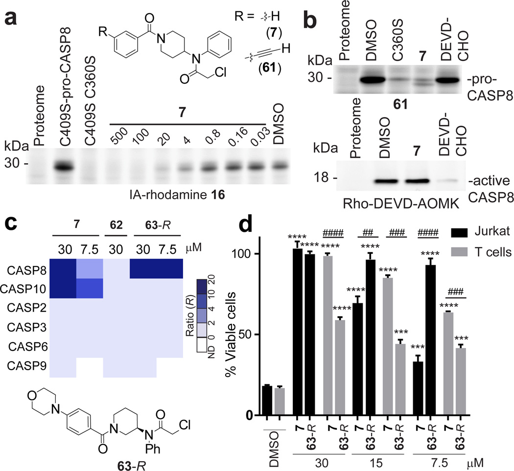
Small molecules are powerful tools for investigating protein function and can serve as leads for new therapeutics. Most human proteins, however, lack small-molecule ligands, and entire protein classes are considered 'undruggable'. Fragment-based ligand discovery can identify small-molecule probes for proteins that have proven difficult to target using high-throughput screening of complex compound libraries. Although reversibly binding ligands are commonly pursued, covalent fragments provide an alternative route to small-molecule probes, including those that can access regions of proteins that are difficult to target through binding affinity alone. Here we report a quantitative analysis of cysteine-reactive small-molecule fragments screened against thousands of proteins in human proteomes and cells. Covalent ligands were identified for >700 cysteines found in both druggable proteins and proteins deficient in chemical probes, including transcription factors, adaptor/scaffolding proteins, and uncharacterized proteins. Among the atypical ligand-protein interactions discovered were compounds that react preferentially with pro- (inactive) caspases. We used these ligands to distinguish extrinsic apoptosis pathways in human cell lines versus primary human T cells, showing that the former is largely mediated by caspase-8 while the latter depends on both caspase-8 and -10. Fragment-based covalent ligand discovery provides a greatly expanded portrait of the ligandable proteome and furnishes compounds that can illuminate protein functions in native biological systems.


Figures

Extended Data Figure 1
Composition of fragment electrophile library and structures of additional tool compounds, click probes, and fragments.
Extended Data Figure 2
Extended Data Figure 2. Analysis of proteomic reactivities of fragment electrophiles in human cell lysates
a, Initial analysis of the proteomic reactivity of fragments using an IA-rhodamine probe 16. Soluble proteome from Ramos cells was treated with the indicated fragments (500 µM each) for 1 h, followed by labeling with IA-rhodamine (1 µM, 1 h) and analysis by SDS-PAGE and in-gel fluorescence scanning. Several proteins were identified that show impaired reactivity with IA-rhodamine in the presence of one or more fragments (asterisks). Fluorescent gel shown in grayscale. b, Frequency of quantification of all cysteines across the complete set of competitive isoTOP-ABPP experiments performed with fragment electrophiles. Note that cysteines were required to have been quantified in at least three isoTOP-ABPP data sets for interpretation. c, Rank order of proteomic reactivity values (or liganded cysteine rates) of fragments calculated as the percentage of all quantified cysteines with R values ≥ 4 for each fragment. The majority of fragments were evaluated in 2–4 replicate experiments in MDA-MB-231 and/or Ramos cell lysates, and their proteomic reactivity values are reported as mean ± SEM values for the replicates. d, Comparison of the proteomic reactivities of representative fragments screened at 500 versus 25 µM in cell lysates. e, Comparison of proteomic reactivity values for fragments tested in both Ramos and MDA-MB-231 lysates. f, Proteomic reactivity values of representative fragments. g, Relative GSH reactivity for representative fragment electrophiles. Consumption of GSH (125 µM) was measured using Ellman’s reagent (5 mM) after 1 h incubation with the indicated fragments (500 µM). h, Proteomic reactivity values for fragments electrophiles (500 µM) possessing different electrophilic groups attached to a common binding element. i, Concentration-dependent labeling of MDA-MB-231 soluble proteomes with acrylamide 18 and chloroacetamide 19 click probes detected by CuACC with a rhodamine-azide tag and analysis by SDS-PAGE and in-gel fluorescence scanning. For panels f and g, data represent mean values ± SEM for at least three independent experiments.
Extended Data Figure 3
Extended Data Figure 3. Analysis of cysteines liganded by fragment electrophiles in competitive isoTOP-ABPP experiments
a, Representative MS1 ion chromatograms for peptides containing C481 of BTK and C131 of MAP2K7, two cysteines known to be targeted by the anti-cancer drug ibrutinib. Ramos cells were treated with ibrutinib (1 µM, 1 h, red trace) or DMSO (blue trace) and evaluated by isoTOP-ABPP. b, Total number of liganded cysteines found in the active sites and non-active sites of enzymes for which X-ray and/or NMR structures have been reported (or reported for a close homologue of the enzyme). c, Functional categorization of liganded and unliganded cysteines based on residue annotations from the Uniprot database. d, Number of liganded and quantified cysteines per protein measured by isoTOP-ABPP. Respective average values of one and three for liganded and quantified cysteines per protein were measured by isoTOP-ABPP. e R values for six cysteines in XPO1 quantified by isoTOP-ABPP, identifying C528 as the most liganded cysteine in this protein. Each point represents a distinct fragment-cysteine interaction quantified by isoTOP-ABPP. f-h Histograms depicting the percentage of fragments that are hits (R ≥ 4) for all 768 liganded cysteines (f), for liganded cysteines found in enzymes for which X-ray and/or NMR structures have been reported (or reported for a close homologue of the enzyme) (g), or for active- and non-active site cysteines in kinases (h). i, Percentage of liganded cysteines targeted only by group A (red) or B (blue) fragments or both group A and B fragments (black). Shown for all liganded cysteines, liganded cysteines in enzyme active and non-active sites, and liganded cysteines in transcription factors/ regulators. For g, i, active-site cysteines were defined as those that reside < 10 Å from established active-site residues and/or bound substrates/inhibitors in enzyme structures. j, The percentage of liganded cysteines in kinases that were targeted by only group A, only group B, or both group A and B compounds. k, Heatmap showing representative fragment interactions for liganded cysteines found in the active sites and non-active sites of kinases. l, Heatmap showing representative fragment interactions for liganded cysteines found in transcription factors/regulators. m, The fraction of liganded (62%; 341 of 553 quantified cysteines) and unliganded (20%; 561 of 2870 quantified cysteines) cysteines that are sensitive to heat denaturation measured by IA-alkyne labeling (R > 3 native/heat denatured). n, Percentage of proteins identified by isoTOP-ABPP as liganded by fragments 3 and 14 and enriched by their corresponding click probes 19 and 18 that are sensitive to heat denaturation (64% (85 of 133 quantified protein targets) and 73% (19 of 26 quantified protein targets), respectively). Protein enrichment by 18 and 19 was measured by whole protein capture of isotopically-SILAC labeled MDA-MB-231 cells using quantitative (SILAC) proteomics. o, The fraction of cysteines predicted to be ligandable or unligandable by reactive docking that were quantified in isoTOP-ABPP experiments. p, The fraction of cysteines predicted to be ligandable or unligandable by reactive docking that show heat-sensitive labeling by the IA-alkyne probe (R > 3 native/heat denatured).
Extended Data Figure 4
Extended Data Figure 4. Confirmation and functional analysis of fragment-cysteine interactions in PRMT1 and MLTK
a, Representative MS1 chromatograms for the indicated Cys-containing peptides from PRMT1 quantified in competitive isoTOP-ABPP experiments of MDA-MB-231 cell lysates, showing blockade of IA-alkyne 1 labeling of C109 by fragment 11, but not control fragment 3. b, 11, but not 3 blocked IA-rhodamine (2 µM) labeling of recombinant, purified WT-PRMT1 (1 µM protein doped into HEK293T cell lysates). Note that a C109S–PRMT1 mutant did not react with IA-rhodamine. c, Apparent IC50 curve for blockade of 16 labeling of PRMT1 by 11. CI, 95% confidence intervals. d, Effect of 11 and control fragment 3 on methylation of recombinant histone 4 by recombinant PRMT1. Shown is one representative experiment of three independent experiments that yielded similar results. e, Representative MS1 ion chromatograms for the MLTK tryptic peptide containing liganded cysteine C22 quantified by isoTOP-ABPP in MDA-MB-231 lysates treated with fragment 4 or control fragment 3 (500 µM each). f, 60, but not control fragment 3 (50 µM of each fragment) blocked labeling of recombinant MLTK (or ZAK) kinase by a previously reported ibrutinib-derived activity probe 59 (upper panel). A C22A–MLTK mutant did not react with the ibrutinib probe. Anti-FLAG blotting confirmed similar expression of WT- and C22A–MLTK proteins, which were expressed as FLAG-fusion proteins in HEK293T cells (lower panel). g, Lysates from HEK293T cells expressing WT- or C22A–MLTK treated with the indicated fragments and then an ibrutinib-derived activity probe 59 at 10 µM . MLTK labeling by 59 was detected by CuAAC conjugation to a rhodamine-azide tag and analysis by SDS-PAGE and in-gel fluorescence scanning. h, Apparent IC50 curve for blockade of ibrutinib probe-labeling of MLTK by 60. i, 60, but not control fragment 3 (100 µM of each fragment) inhibited the kinase activity of WT-, but not C22A–MLTK. For panels c, h and i, data represent mean values ± SEM for at least three independent experiments. Statistical significance was calculated with unpaired students t-tests comparing DMSO- to fragment-treated samples; **, p < 0.1.
Extended Data Figure 5
Extended Data Figure 5. Confirmation and functional analysis of fragment-cysteine interactions in IMPDH2 and TIGAR
a, Representative MS1 ion chromatograms for IMPDH2 tryptic peptides containing the catalytic cysteine, C331, and Bateman domain cysteine, C140, quantified by isoTOP-ABPP in cell lysates treated with the indicated fragments (500 µM each). b, Structure of human IMPDH2 (PDB ID: 1NF7) (light grey) and its structurally unresolved Bateman domain modeled by ITASSER55 (dark grey) showing the positions of C331 (red spheres), ribavirin monophosphate and C2-mycophenolic adenine dinucleotide (blue), and C140 (yellow spheres). c, Click probe 18 (25 µM) labeled WT-IMPDH2 and C331S–IMPDH2, but not C140S–IMPDH2 (or C140S/C331S–IMPDH2). Labeling was detected by CuAAC conjugation to a rhodamine-azide reporter tag and analysis by SDS-PAGE and in-gel fluorescence scanning. Recombinant IMPDH2 WT and mutants were expressed and purified from E. coli and added to Jurkat lysates to a final concentration of 1 µM protein. d, Fragment reactivity with recombinant, purified IMPDH2 added to Jurkat lysates to a final concentration of 1 µM protein, where reactivity was detected in competition assays using the click probe 18 (25 µM). Note that 18 reacted with WT- and C331S–IMPDH2, but not C140S or C140S/C331S–IMPDH2. e, Nucleotide competition of 18 (25 µM) labeling of WT-IMPDH2 added to MDA-MB-231 lysates to a final concentration of 1 µM protein. f, Nucleotide competition profile for 18-labeling of recombinant WT-IMPDH2 (500 µM of each nucleotide). g, Apparent IC50 curve for blockade of 18 labeling of IMPDH2 by ATP. h, Representative MS1 chromatograms for TIGAR tryptic peptides containing C114 and C161 quantified by isoTOP-ABPP in cell lysates treated with the indicated fragments (500 µM each). i, Crystal structure of TIGAR (PDB ID: 3DCY) showing C114 (red spheres), C161 (yellow spheres), and inorganic phosphate (blue). j, Labeling of recombinant, purified TIGAR and mutant proteins by the IA-rhodamine (2 µM) probe. TIGAR proteins were added to MDA-MB-231 lysates, to a final concentration of 2 µM protein. k, 5, but not control fragment 3 blocked IA-rhodamine (2 µM) labeling of recombinant, purified C161S–TIGAR (2 µM protein doped into Ramos cell lysates). l, Apparent IC50 curve for blockade of IA-rhodamine labeling of C161S–TIGAR by 5. m, 5, but not control fragment 3 (100 µM of each fragment) inhibited the catalytic activity of WT-TIGAR, C161S–TIGAR, but not C114S–TIGAR or C114S/C161S–TIGAR. n, Concentration-dependent inhibition of WT-TIGAR by 5. Note that the C140S–TIGAR mutant was not inhibited by 5. Data represent mean values ± SEM for 4 replicate experiments at each concentration. For panels f, g and l-n, data represent mean values ± SEM for at least three independent experiments. Statistical significance was calculated with unpaired students t-tests comparing DMSO- to fragment-treated samples; **, p < 0.01, ****, p < 0.0001.
Extended Data Figure 6
Extended Data Figure 6. IDH1-related and general in situ activity of fragment electrophiles
a, X-ray crystal structure of IDH1 (PDB ID: 3MAS) showing the position of C269 and the frequently mutated residue in cancer, R132. b, Blockade of 16 labeling of WT-IDH1 by representative fragment electrophiles. Recombinant, purified WT-IDH1 was added to MDA-MB-231 lysates at a final concentration of 2 µM, treated with fragments at the indicated concentrations, followed by IA-rhodamine probe 16 (2 µM) and analysis by SDS-PAGE and in-gel fluorescence scanning. Note that a C269S mutant of IDH1 did not label with IA-rhodamine 16. c, d, Reactivity of 20 and control fragment 2 with recombinant, purified WT-IDH1 (b) or R132H–IDH1 (c) added to MDA-MB-231 lysates to a final concentration of 2 or 4 µM protein, respectively. Fragment reactivity was detected in competition assays using the IA-rhodamine probe (2 µM); note that the C269S–IDH1 mutant did not react with IA-rhodamine. e, f, Apparent IC50 curve for blockade of IA-rhodamine-labeling of IDH1 (e) and R132H–IDH1 (f) by 20. Note that the control fragment 2 showed much lower activity. g, Representative MS1 ion chromatograms for the IDH1 tryptic peptides containing liganded cysteine C269 and an unliganded cysteine C379 quantified by isoTOP-ABPP in MDA-MB-231 lysates treated with fragment 20 (25 µM). h, 20, but not 2, inhibited IDH1-catalyzed oxidation of isocitrate to α-ketoglutarate (α-KG) as measured by an increase in NADPH production (340 nm absorbance). 20 did not inhibit the C269S–IDH1 mutant. i, 20 inhibited oncometabolite 2-hydroxyglutarate (2-HG) production by R132H–IDH1. MUM2C cells stably overexpressing the oncogenic R132H–IDH1 mutant or control GFP-expressing MUM2C cells were treated with the indicated fragments (2 h, in situ). Cells were harvested, lysed and IDH1-dependent production of 2-HG from α-KG and NADPH was measured by LC-MS and from which 2-HG production of GFP-expressing MUM2C cells was subtracted (GFP-expressing MUM2C cells produced < 10% of the 2-HG generated by R132H–IDH1-expressing MUM2C cells). j, Western blot of MUM2C cells stably overexpressing GFP (mock) or R132H–IDH1 proteins. k, Representative MS1 chromatograms for the IDH1 tryptic peptides containing liganded cysteine C269 and an unliganded cysteine C379 quantified by isoTOP-ABPP in R132H–IDH-expressing MUM2C lysates treated with 20 or control fragment 2 (50 µM, 2 h, in situ). l, Proteomic reactivity values for individual fragments are comparable in vitro and in situ. One fragment (11) marked in red showed notably lower reactivity in situ versus in vitro. Reactivity values were calculated as in Fig. 1c. Dashed line mark 90% prediction intervals for the comparison of in vitro and in situ proteomic reactivity values for fragment electrophiles. Blue and red circles mark fragments that fall above (or just at) or below these prediction intervals, respectively. m, Fraction of cysteines liganded in vitro that are also liganded in situ. Shown are liganded cysteine numbers for individual fragments determined in vitro and the fraction of these cysteines that were liganded by the corresponding fragments in situn, Representative cysteines that were selectively targeted by fragments in situ, but not in vitro. For in situ-restricted fragment-cysteine interactions, a second cysteine in the parent protein was detected with an unchanging ratio (R ~ 1), thus controlling for potential fragment-induced changes in protein expression. For panels e, f, h and i, data represent mean values ± SEM for at least three independent experiments. Statistical significance was calculated with unpaired students t-tests comparing DMSO- to fragment-treated samples; ****, p < 0.0001.
Extended Data Figure 7
Extended Data Figure 7. Fragment electrophiles that target pro-CASP8
a, Representative MS1 chromatograms for CASP8 tryptic peptide containing the catalytic cysteine C360 quantified by isoTOP-ABPP in cell lysates or cells treated with fragment 4 (250 µM, in vitro; 100 µM, in situ) and control fragment 21 (500 µM, in vitro; 200 µM, in situ). b, Neither 7 nor control fragment 62 (100 µM each) inhibited recombinant, purified active CASP3 and CASP8 assayed using DEVD-AMC and IETD-AFC substrates, respectively. DEVD-CHO (20 µM) inhibited both caspases. c, Fragment reactivity with recombinant, purified active CASP8 added to cell lysates, where reactivity was detected in competition assays using the caspase activity probe Rho-DEVD-AOMK probe (2 µM, 1 h). d, Western blot of proteomes from MDA-MB-231, Jurkat, and CASP8-null Jurkat proteomes showing that CASP8 was only found in the pro-enzyme form in these cells. e, Fragment reactivity with recombinant, purified pro-CASP8 (D374A, D384A, C409S) added to cell lysates to a final concentration of 1 µM protein, where reactivity was detected in competition assays with the IA-rhodamine probe (2 µM). Note that mutation of both cysteine-360 and cysteine-409 to serine prevented labeling of pro-CASP8 by IA-rhodamine. f, inactive control fragment 62 did not compete IA-rhodamine labeling of C360 of pro-CASP8. g, Apparent IC50 curve for blockade of IA-rhodamine labeling of pro-CASP8 (C409S) by 7. h, 7 (50 µM) fully competed IA-alkyne-labeling of C360 of endogenous CASP8 in cell lysates as measured by isoTOP-ABPP. Representative MS1 chromatograms are shown for the C360-containing peptide of CASP8. i, Concentration-dependent reactivity of click probe 61, with recombinant, purified pro-CASP8 (D374A, D384A) added to cell lysates to a final concentration of 1 µM protein. Note that pre-treatment with 7 blocked 61 reactivity with pro-CASP8 and mutation of C360 to serine prevented labeling of pro-CASP8 by 61 (25 µM). ). j, 7 (30 µM) blocked IA-alkyne labeling of C360 of pro-CASP8, but not active-CASP8 as measured by isoTOP-ABPP. Recombinant pro- and active-CASP8 were added to Ramos lysates at 1 µM and then treated with 7 (30 µM) followed by isoTOP-ABPP. k, Fragments 7 and 62 did not block labeling by Rho-DEVD-AOMK (2 µM) of recombinant, purified active-CASP8 and active-CASP3 added to MDA-MB-231 cell lysates to a final concentration of 1 µM protein. l, 7 does not inhibit active caspases. Recombinant, active caspases were added to MDA-MD-231 lysate to a final concentration of 200 nM (CASP2, 3, 6, 7) or 1 µM (CASP8, 10), treated with z- VAD-FMK (25 µM) or 7 (50 µM), followed by labeling with the Rho-DEVD-AOMK probe (2 µM). m, Representative MS1 chromatograms for tryptic peptides containing the catalytic cysteines of CASP8 (C360), CASP2 (C320), and CASP7 (C186) quantified by isoTOP-ABPP in Jurkat cell lysates treated with 7 or 62 (50 µM, 1 h). n, 7, but not control fragment 62, blocked extrinsic, but not intrinsic apoptosis. Jurkat cells (1.5 million cells/mL) were incubated with 7 or 62 (30 µM) or the pan-caspase inhibitor VAD-FMK (100 µM) for 30 min prior to addition of staurosporine (2 µM) or SuperFasLigand™ (100 ng/mL). Cells were incubated for 4 h and viability was quantified with CellTiter-Glo®. RLU- relative light unit. o, For cells treated as described in n, cleavage of PARP (96 kDa), CASP8 (p43/p41, p18), and CASP3 (p17) was visualized by western blot. p, 7 protects Jurkat cells from extrinsic, but not intrinsic apoptosis. Cleavage of PARP, CASP8, and CASP3 detected by western blotting as shown in panel o was quantified for three (STS) or two (FasL) independent experiments. Cleavage products (PARP (96 kDa), CASP8 (p43/p41), CASP3 (p17)) were quantified for compound treatment and the % cleavage relative to DMSO-treated samples was calculated. For panels b, g and n, data represent mean values ± SEM for at least three independent experiments. For panel p, STS data represent mean values ± SEM for three independent experiments, and FasL data represent mean values ± SD for two independent experiments. Statistical significance was calculated with unpaired students t-tests comparing active compounds (VAD-FMK and 7) to control compound 62; **, p < 0.01, ***, p < 0.001, ****, p < 0.0001.
Extended Data Figure 8
Extended Data Figure 8. CASP10 is involved in intrinsic apoptosis in primary human T cells
a, Representative MS1 peptide signals showing R values for caspases detected by quantitative proteomics using probe 61. ABPP-SILAC experiments. Jurkat cells (10 million cells) were treated with either DMSO (heavy cells) or the indicated compounds (light cells) for 2 h followed by probe 61 (10 µM, 1 h). b, 7 competed 61-labeling of pro-CASP8 and CASP10, whereas 63-R selectively blocked probe labeling of pro-CASP8. c, 7, but not 63-R block probe labeling of pro-CASP10. Recombinant pro-CASP10 was added to MDA-MB-231 lysates to a final concentration of 300 nM, treated with the indicated compounds, and labeled with probe 61. Mutation of the catalytic cysteine C401A fully prevented labeling by 61. d, Apparent IC50 curve for blockade of 61-labeling of pro-CASP10 by 7, 63-R or 63-Se, Neither 7 nor 63 (25 µM each) inhibited the activity of recombinant, purified active CASP10 (500 nM), which was assayed following addition of the protein to MDA-MB-231 lysate using fluorometric AEVD-AMC substrate. DEVD-CHO (20 µM) inhibited the activity of CASP10. f, Apparent IC50 curve for blockade of 61 labeling of pro-CASP8 and pro-CASP10 by 63-Rg, 63-R shows increased potency against pro-CASP8. Recombinant pro-CASP8 was added to MDA-MB-231 lysates to a final concentration of 300 nM, treated with the indicated compounds and labeled with probe 61. h, Apparent IC50 curve for blockade of 61 labeling of pro-CASP8 by 63R compared with 63- S. The structure of 63-S is shown. i, CASP10 is more highly expressed in primary human T cells compared to Jurkat cells. Western blot analysis of full-length CASP10, CASP8 and GAPDH expression levels in Jurkat and T-cell lysates (2 mg/mL). j, Jurkat cells (150,000 cells/well) were incubated with 7 or 63R at the indicated concentrations for 30 min prior to addition of staurosporine (2 µM) or SuperFasLigand™ (100 ng /mL). Cells were incubated for 4 h and viability was quantified with CellTiter-Glo®. k, Jurkat cells treated as in j, but with 63-Ror 63-Sl, HeLa cells (20,000 cells/well) were seeded and 24 h later treated with the indicated compounds for 30 minutes prior to addition of SuperFasLigand™ (100 ng /mL) and cycloheximide (CHX, 2.5 ng/mL). Cells were incubated for 6 h and viability quantified with CTG. m, For T cells treated as in Fig. 4d cleavage of CASP10 (p22), CASP8 (p18), CASP3 (p17) and RIPK (33 kDa) was visualized by western blotting. For panels d-f, h, and j-k, data represent mean values ± SEM for at least three independent experiments. Statistical significance was calculated with unpaired students t-tests comparing DMSO- to fragment-treated samples; **, p < 0.01, ****, p < 0.0001.
Figure 1
Figure 1. Proteome-wide screening of covalent fragments
a, General protocol for competitive isoTOP-ABPP. Competition ratios, or R values, are measured by dividing the MS1 ion peaks for IA-alkyne (1)-labeled peptides in DMSO-treated (heavy, or blue) versus fragment-treated (light, or red) samples. b, General structure of electrophilic fragment library, where the reactive (electrophilic) and binding groups are colored green and black, respectively. c, Competitive isoTOP-ABPP analysis of the MDA-MB-231 cell proteome pre-treated with the electrophilic 3,5-di(trifluoromethyl)aniline chloroacetamide 3 and acrylamide 14 fragments, along with the non-electrophilic acetamide analogue 17 (500 µM each). Proteomic reactivity values, or liganded cysteine rates, for fragments were calculated as the percentage of total cysteines with R values ≥ 4 in DMSO/fragment (heavy/light) comparisons. d, Representative MS1 peptide ion chromatograms from competitive isoTOP-ABPP experiments marking liganded cysteines selectively targeted by one of three fragments 3, 4, and 23.
Figure 2
Figure 2. Analysis of cysteines and proteins liganded by fragment electrophiles
a, Fraction of total quantified cysteines and proteins that were liganded by fragment electrophiles in competitive isoTOP-ABPP experiments. b, Fraction of liganded proteins found in DrugBank. c, Functional classes of DrugBank and non-DrugBank proteins containing liganded cysteines. d, Comparison of the ligandability of cysteines as a function of their intrinsic reactivity with the IA-alkyne probe. Cysteine reactivity values (left y-axis) were taken from reference, where lower ratios correspond to higher cysteine reactivity. A moving average with a step-size of 50 is shown in blue for the percentage of liganded cysteines within each reactivity bin (percent values shown on right y-axis).
Figure 3
Figure 3. Analysis of fragment-cysteine interactions
a, Heatmap showing R values for representative cysteines and fragments organized by proteomic reactivity values (high to low, left to right) and percentage of fragment hits for individual cysteines (high to low, top to bottom). R values ≥ 4 designate fragment hits (colored medium and dark blue). White color – not detected (ND). b, Representative example of reactive docking predictions shown for XPO1 (PDB ID: 3GB8). All accessible cysteines were identified and reactive docking was conducted with all fragments from the library within a 25 Å docking cube centered on each accessible cysteine (cube shown in green for liganded Cys in XPO1; see Supplementary Information for more details). Legend presents categories of XPO1 cysteines based on combined docking and isoTOP-ABPP results. c, Success rate of reactive docking predictions for liganded cysteines identified by isoTOP-ABPP for 29 representative proteins.
Figure 4
Figure 4. Electrophile compounds that target pro-CASP8 and pro-CASP10
a, Compound 7 blocked 16 labeling of recombinant, purified pro-CASP8 (bearing a C409S mutation to eliminate IA-rhodamine labeling at this site; added to Ramos cell lysate at 1 µM). Note that a C360S/C409S–mutant of pro-CASP8 did not label with 16. b, 7 blocked probe labeling of pro-, but not active CASP8. Recombinant pro- and active- CASP8 (1 µM) were treated with 7 (50 µM) or Ac-DEVD-CHO (20 µM), for 1 h followed by click probe 61 (25 µM) for pro-CASP8 and the Rho-DEVD-AOMK probe (2 µM) for active-CASP8. c, Heatmap showing R values for caspases measured by quantitative proteomics in Jurkat cells treated with 7, 63R, or 62 followed by probe 61 (10 µM, 1 h). d, Comparison of effects of 7 and 63R on FasL-induced apoptosis in Jurkat cells or anti-CD3, anti-CD28-activated primary human T cells. For d, data represent mean values ± SEM for at least three independent experiments, and results are representative of multiple experiments performed with T cells from different human subjects. Statistical significance was calculated with unpaired students t-tests comparing DMSO- to fragment-treated samples; ****, p < 0.0001 and comparing Jurkat to T cells ####, p < 0.0001.

References

    1. Edfeldt FN, Folmer RH, Breeze AL. Fragment screening to predict druggability (ligandability) and lead discovery success. Drug discovery today. 2011;16:284–287. - PubMed
    1. Hopkins AL, Groom CR. The druggable genome. Nature reviews. Drug discovery. 2002;1:727–730. - PubMed
    1. Scott DE, Coyne AG, Hudson SA, Abell C. Fragment-based approaches in drug discovery and chemical biology. Biochemistry. 2012;51:4990–5003. - PubMed
    1. Erlanson DA, et al. Site-directed ligand discovery. Proceedings of the National Academy of Sciences of the United States of America. 2000;97:9367–9372. - PMC - PubMed
    1. Cardoso R, et al. Identification of Cys255 in HIF-1alpha as a novel site for development of covalent inhibitors of HIF-1alpha/ARNT PasB domain protein-protein interaction. Protein science : a publication of the Protein Society. 2012;21:1885–1896. - PMC - PubMed

References for online-only Methods

    1. Weerapana E, Speers AE, Cravatt BF. Tandem orthogonal proteolysis-activity-based protein profiling (TOP-ABPP)--a general method for mapping sites of probe modification in proteomes. Nat Protoc. 2007;2:1414–1425. - PubMed
    1. Inloes JM, et al. The hereditary spastic paraplegia-related enzyme DDHD2 is a principal brain triglyceride lipase. Proceedings of the National Academy of Sciences of the United States of America. 2014;111:14924–14929. - PMC - PubMed
    1. Adibekian A, et al. Click-generated triazole ureas as ultrapotent in vivo-active serine hydrolase inhibitors. Nature Chemical Biology. 2011;7:469–478. - PMC - PubMed
    1. Wang C, Weerapana E, Blewett MM, Cravatt BF. A chemoproteomic platform to quantitatively map targets of lipid-derived electrophiles. Nat Meth. 2014;11:79–85. - PMC - PubMed
    1. Consortium TU. UniProt: a hub for protein information. Nucleic Acids Research. 2015;43:D204–D212. - PMC - PubMed

Publication types

MeSH terms