. 2016 Jul 7;535(7610):136-9.
doi: 10.1038/nature18010. Epub 2016 Jun 15.

Design of a hyperstable 60-subunit protein dodecahedron. [corrected]

Design of a hyperstable 60-subunit protein dodecahedron. [corrected]

Yang Hsia et al. Nature. .

Erratum in

  • Corrigendum: Design of a hyperstable 60-subunit protein icosahedron.
    Hsia Y, Bale JB, Gonen S, Shi D, Sheffler W, Fong KK, Nattermann U, Xu C, Huang PS, Ravichandran R, Yi S, Davis TN, Gonen T, King NP, Baker D. Hsia Y, et al. Nature. 2016 Dec 1;540(7631):150. doi: 10.1038/nature20108. Epub 2016 Oct 19. Nature. 2016. PMID: 27760110 No abstract available.

Abstract

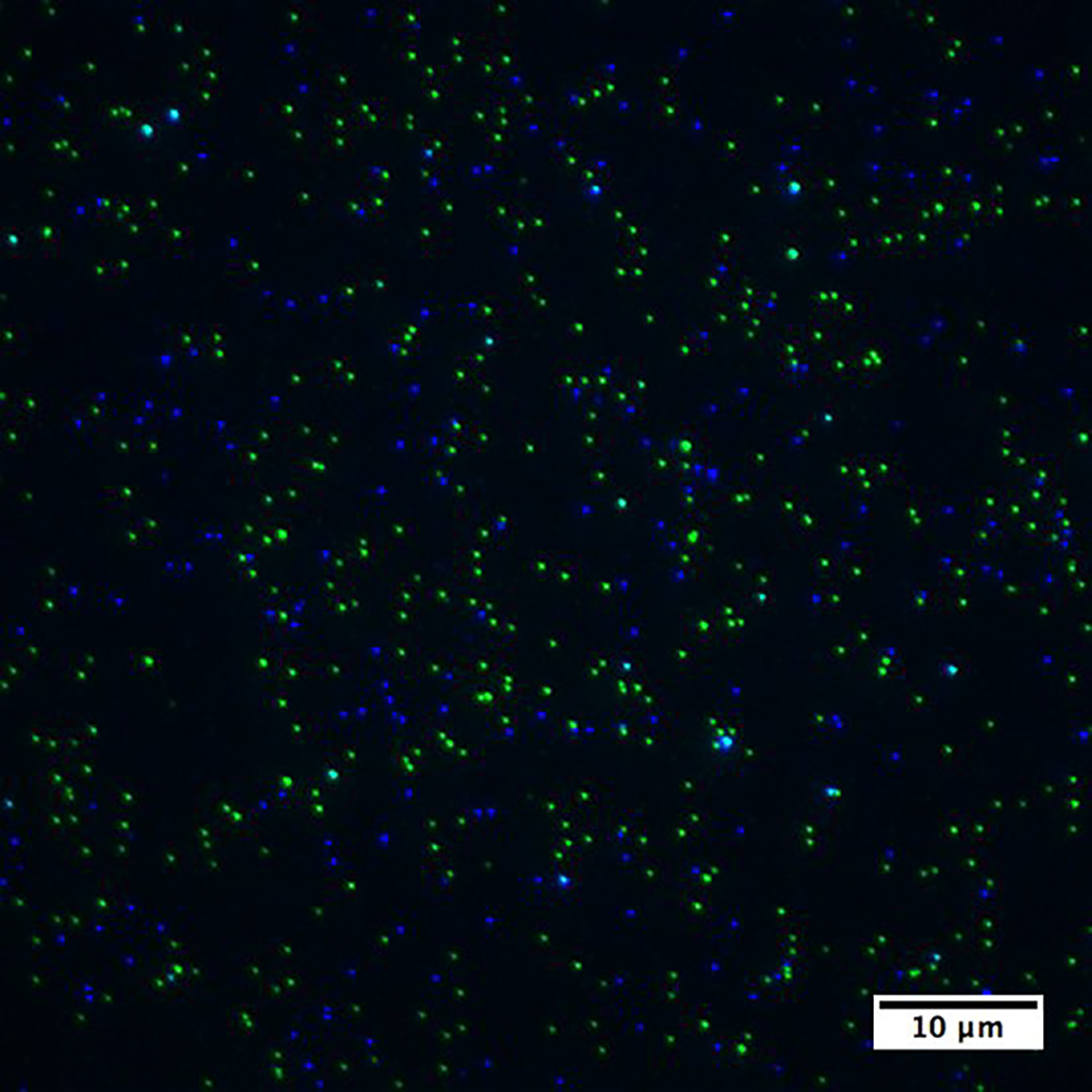
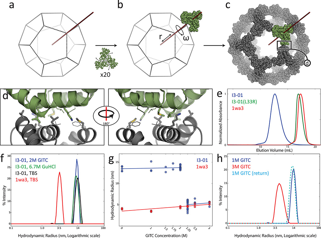
The dodecahedron [corrected] is the largest of the Platonic solids, and icosahedral protein structures are widely used in biological systems for packaging and transport. There has been considerable interest in repurposing such structures for applications ranging from targeted delivery to multivalent immunogen presentation. The ability to design proteins that self-assemble into precisely specified, highly ordered icosahedral structures would open the door to a new generation of protein containers with properties custom-tailored to specific applications. Here we describe the computational design of a 25-nanometre icosahedral nanocage that self-assembles from trimeric protein building blocks. The designed protein was produced in Escherichia coli, and found by electron microscopy to assemble into a homogenous population of icosahedral particles nearly identical to the design model. The particles are stable in 6.7 molar guanidine hydrochloride at up to 80 degrees Celsius, and undergo extremely abrupt, but reversible, disassembly between 2 molar and 2.25 molar guanidinium thiocyanate. The dodecahedron [corrected] is robust to genetic fusions: one or two copies of green fluorescent protein (GFP) can be fused to each of the 60 subunits to create highly fluorescent ‘standard candles’ for use in light microscopy, and a designed protein pentamer can be placed in the centre of each of the 20 pentameric faces to modulate the size of the entrance/exit channels of the cage. Such robust and customizable nanocages should have considerable utility in targeted drug delivery, vaccine design and synthetic biology.


Figures

Extended Data Figure 1
Extended Data Figure 1. I3-01 Tolerance to Temperature
Dynamic light scattering measurements as I3-01 is subjected to heating to 90°C (solid line), then cooling to 25°C (dotted line) in a) TBS, b) 6.7 M GuHCl, and c) 2 M GITC. In all 3 conditions, any indications of aggregation or increase in size due to temperature appear to be completely reversible.
Extended Data Figure 2
Extended Data Figure 2. Reproducibility of I3-01 transition in 2 to 2.25 M GITC
Four examples each of independent measurements at 2 M (blue) and 2.25 M (red) GITC using DLS show the reproducibility of the cage disassociation. Histograms are plotted offset by 1% intensity from each other for clarity.
Extended Data Figure 3
Extended Data Figure 3. SEC of T33-21 and I3-01 fused with sfGFP
Size exclusion chromatography traces for T33-21 (12-mer in red and 24-mer in blue) and I3-01 (60-mer in green and 120-mer in purple) sfGFP fusions, display increased particle sizes with increasing copies of GFP, but retain monodispersed populations. The N-terminal fusion of sfGFP (dashed line) is expected to extend mostly outwards of the icosahedron, thus greatly increasing the hydrodynamic radius while the C-terminal fusion is predicted to occupy the internal void space.
Extended Data Figure 4
Extended Data Figure 4. Tolerance of I3-01-sfGFP fusions to GuHCl
N-terminal (red) and C-terminal (blue) sfGFP fusions were equilibrated to 0–6.4 M GuHCl. UV absorbance at 490 nm monitors the unfolding of sfGFP (top, solid line). DLS experiments (top, dotted line) reveal as sfGFP unfolds, the hydrodynamic radius increases slightly, and then stabilizes. Bottom panels show that in 1 M GuHCl (solid line) and in 6 M GuHCl (dotted line), the icosahedral assemblies remain relatively monodisperse.
Extended Data Figure 5
Extended Data Figure 5. I3-01 C-terminal fusions with other fluorescent proteins
Fluorescent proteins mTurquoise2 or sYFP2 were fused to the C-terminus of I3-01. The field of view using widefield fluorescence microscopy shows distinct signals of each type when the two types are mixed together.
Extended Data Figure 6
Extended Data Figure 6. I3-01 Retains Native Enzyme Activity
Coupled KDPG aldolase assay showing native-like enzymatic activity in I3-01. The K129A knockout shows no enzyme activity, similar to buffer alone.
Figure 1
Figure 1. Design methodology and biochemical characterization
a–b) Icosahedral 3-fold axis in red and aligned trimeric building block in green. c) Optimization of r and ω yields closely opposed interfaces between subunits. d) Sequence design yields low energy interfaces; in the I3-01 case, composed of 5 designed residues (thick) and 2 native residues (thin). e) I3-01 appears larger by SEC than the similarly sized I3-01(L33R) and wild type trimer (1wa3). f) DLS measurement of hydrodynamic radius of 1wa3 (3.5 nm) and I3-01 (14 nm). I3-01 remains assembled in 6.7 M GuHCl and in 2 M GITC. g) Extremely sharp disassociation to trimeric building blocks at 2.25 M GITC. Data points represent independent measurements. h) I3-01 icosahedron disassembles into the trimeric building blocks at 3 M GITC, and reassembles following dilution to 1 M.
Figure 2
Figure 2. Cryo-Electron Microscopy
a) Field of view cryo-EM micrograph with homogeneous icosahedral particles in various orientations. b) Back projections of I3-01 from the design model. c) CryoEM class averages closely match the design projections along all three symmetry axes. d–e) The calculated density (blue, 3.22 σ) closely matches the design model (green).
Figure 3
Figure 3. Tuning nanocage structure and function with genetic fusions
a) (left) Cryo-EM micrograph of I3-01(ctGFP). (top right) Computational model; sfGFP in green. (bottom right) Class average along the 5-fold axis. b) Fluorescence microscopy fields of view, c) fluorescence intensity histograms, and d) correlation between the mean fluorescence intensity and sfGFP copy number for nanoparticles with different numbers of fused sfGFP molecules. e–f) Computational model and class averages along the five fold axis of negatively stained e) I3-01 and f) I3-01(HB); helical bundle in red. Three class averages are shown as there is some heterogeneity at the pentameric face. Weak density in the center of the pentameric faces in I3-01 may reflect randomly packaged material. There is clear density in the center of the pentameric faces in the I3-01(HB) class averages consistent with the model.

References

    1. Zandi R, Reguera D, Bruinsma RF, Gelbart WM, Rudnick J. From The Cover: Origin of icosahedral symmetry in viruses. Proc. Natl. Acad. Sci. 2004;101:15556–15560. - PMC - PubMed
    1. Ritsert K, et al. Studies on the lumazine synthase/riboflavin synthase complex of Bacillus subtilis: crystal structure analysis of reconstituted, icosahedral beta-subunit capsids with bound substrate analogue inhibitor at 2.4 A resolution. J. Mol. Biol. 1995;253:151–167. - PubMed
    1. Howorka S. Rationally engineering natural protein assemblies in nanobiotechnology. Curr. Opin. Biotechnol. 2011;22:485–491. - PubMed
    1. Roldão A, Mellado MCM, Castilho LR, Carrondo MJT, Alves PM. Virus-like particles in vaccine development. Expert Rev. Vaccines. 2010;9:1149–1176. - PubMed
    1. Effio CL, Hubbuch J. Next generation vaccines and vectors: Designing downstream processes for recombinant protein-based virus-like particles. Biotechnol. J. 2015;10:715–727. - PubMed

References for online-only Methods

    1. Zhou Z, et al. Genetically encoded short peptide tags for orthogonal protein labeling by Sfp and AcpS phosphopantetheinyl transferases. ACS Chem. Biol. 2007;2:337–346. - PubMed
    1. Baalousha M, Lead JR. Nanoparticle dispersity in toxicology. Nat. Nanotechnol. 2013;8:308–309. - PubMed
    1. Zulauf M, D’Arcy A. Light scattering of proteins as a criterion for crystallization. J. Cryst. Growth. 1992;122:102–106.
    1. Nannenga BL, Iadanza MG, Vollmar BS, Gonen T. In: Current Protocols in Protein Science. Coligan JE, Dunn BM, Speicher DW, Wingfield PT, editors. John Wiley & Sons Inc.; 2013.
    1. Tang G, et al. EMAN2: an extensible image processing suite for electron microscopy. J. Struct. Biol. 2007;157:38–46. - PubMed

Publication types

MeSH terms

Substances