. 2016 Jun 23;534(7608):562-5.
doi: 10.1038/nature18595. Epub 2016 Jun 15.

Molecular architecture of the human sperm IZUMO1 and egg JUNO fertilization complex

Affiliations

Molecular architecture of the human sperm IZUMO1 and egg JUNO fertilization complex

Halil Aydin et al. Nature. .

Abstract

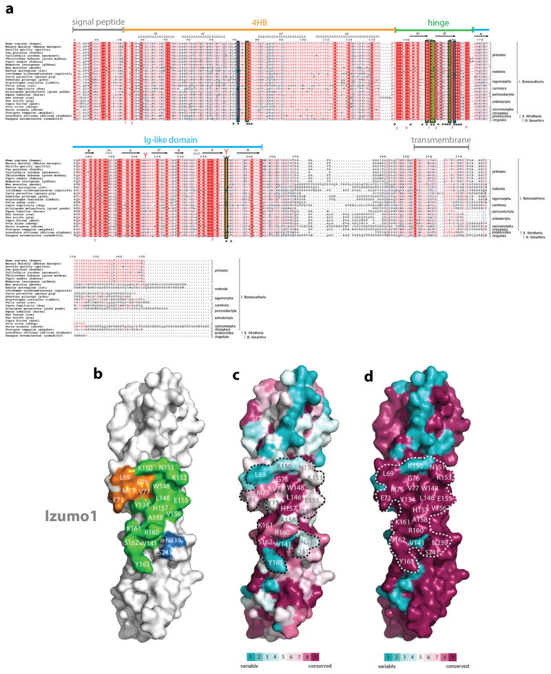
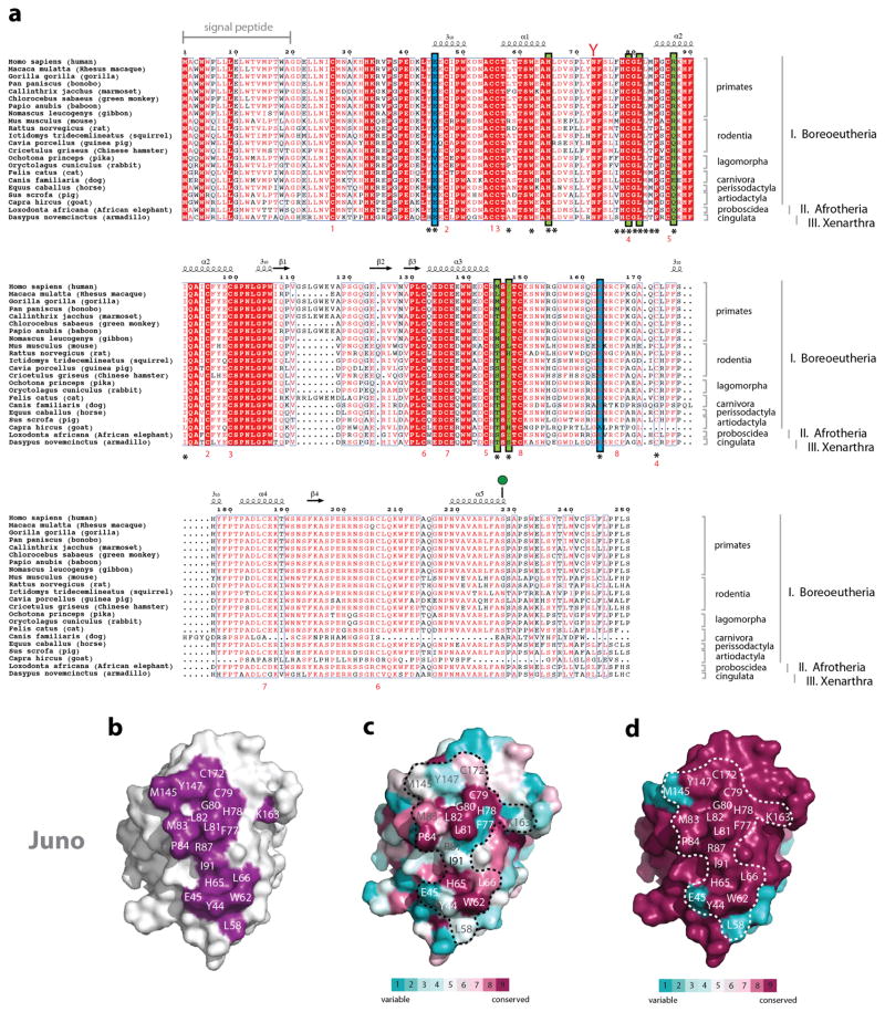
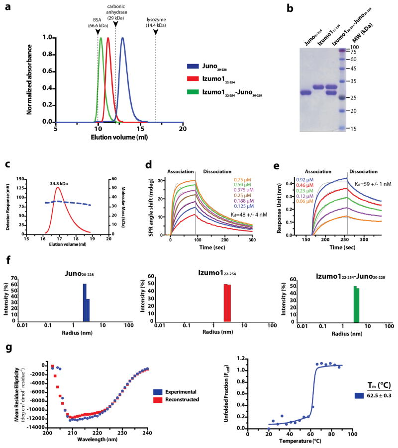
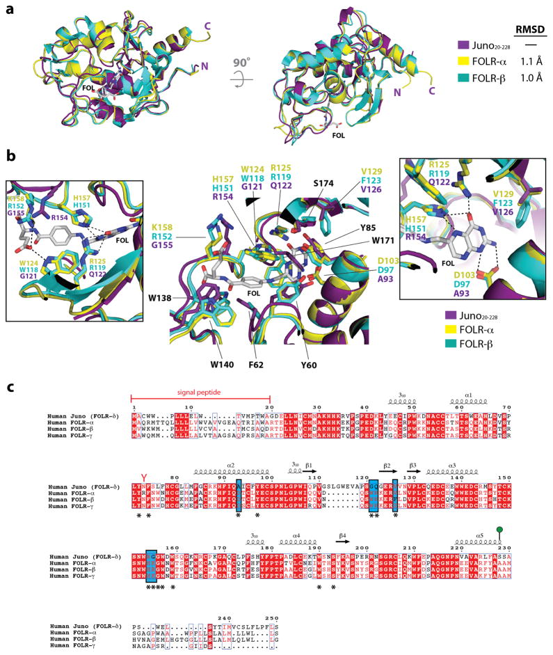
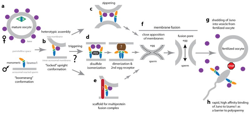
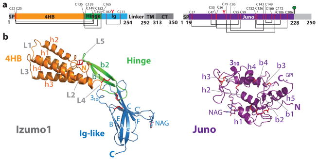
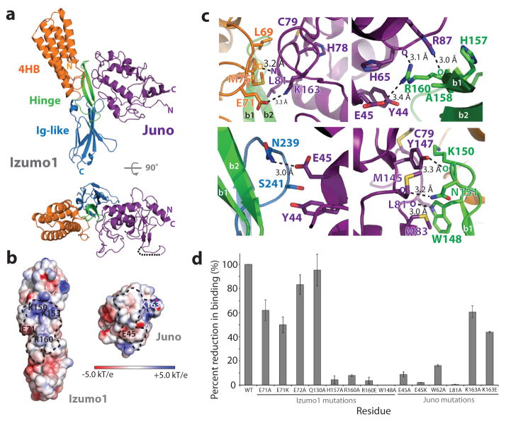
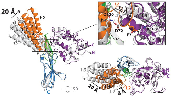
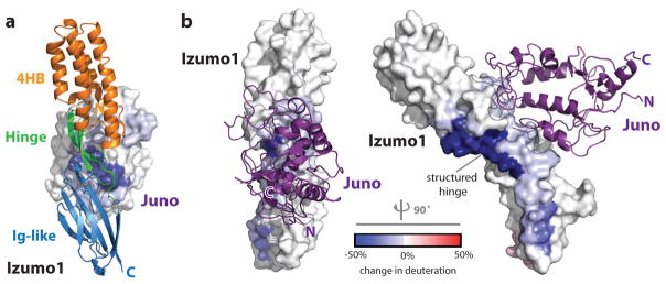
Fertilization is an essential biological process in sexual reproduction and comprises a series of molecular interactions between the sperm and egg. The fusion of the haploid spermatozoon and oocyte is the culminating event in mammalian fertilization, enabling the creation of a new, genetically distinct diploid organism. The merger of two gametes is achieved through a two-step mechanism in which the sperm protein IZUMO1 on the equatorial segment of the acrosome-reacted sperm recognizes its receptor, JUNO, on the egg surface. This recognition is followed by the fusion of the two plasma membranes. IZUMO1 and JUNO proteins are indispensable for fertilization, as constitutive knockdown of either protein results in mice that are healthy but infertile. Despite their central importance in reproductive medicine, the molecular architectures of these proteins and the details of their functional roles in fertilization are not known. Here we present the crystal structures of human IZUMO1 and JUNO in unbound and bound conformations. The human IZUMO1 structure exhibits a distinct boomerang shape and provides structural insights into the IZUMO family of proteins. Human IZUMO1 forms a high-affinity complex with JUNO and undergoes a major conformational change within its N-terminal domain upon binding to the egg-surface receptor. Our results provide insights into the molecular basis of sperm-egg recognition, cross-species fertilization, and the barrier to polyspermy, thereby promising benefits for the rational development of non-hormonal contraceptives and fertility treatments for humans and other mammals.


Figures

Extended Data Figure 1
Extended Data Figure 1. Conservation of Izumo1 residues
(a) Alignment of Izumo1 protein sequences from various mammals. Izumo1 sequences from Homo sapiens (human; GenBank: BAD91012.1), Macaca mulatta (rhesus macaque; GenBank: EHH30233.1), Gorilla gorilla (gorilla; Uniprot: G3QFY5), Pan paniscus (bonobo; NCBI: XP_003814124.1), Callinthrix jacchus (marmoset; Uniprot: F7H859), Chlorocebus sabaeus (green monkey; Uniprot: A0A0D9S2Z4), Papio anubis (baboon; Uniprot: A0A0A0MU86), Nomascus leucogenys (gibbon; Uniprot: G1QXF7), Mus musculus (mouse; GenBank: BAD91011.1), Rattus norvegicus (rat; GenBank: BAD91013.1), Ictidomys tridecemlineatus (squirrel; Uniprot: I3N2L9), Cavia porcellus (guinea pig; Uniprot: H0UTJ7), Ochotona princeps (pika; NCBI: XP_004597241.1), Oryctolagus cuniculus (rabbit; Uniprot: G1TVX5), Felis catus (cat; NCBI: XP_006941089.1), Canis familiaris (dog, Uniprot: F6UM65), Ailuropoda melanoleuca (giant panda, Uniprot: G1M882), Equus caballus (horse; Uniprot: F6YE25), Bos taurus (cow; Uniprot: E1BDA8), Sus scrofa (pig; Uniprot: F1RIQ7), Capra hircus (goat; Uniprot: C6ZEA2), Ovis aries (sheep; Uniprot: W5PRD0), Sorex araneus (shrew; NCBI: XP_004619786.1), Pteropus vampyrus (megabat; NCBI: XP_011372928.1), Loxodonta africana (African elephant; NCBI: XP_003406572.1), and Dasypus novemcinctus (armadillo; NCBI: XP_004451154.1) are aligned. Red boxes indicate complete conservation of a given amino acid. N-linked glycosylation sequons (N-x-S/T) are indicated by red-coloured Y-shaped symbols. Secondary structural elements observed in the crystal structure of Izumo1 are shown as arrows for β-strands and coils for α-helices. Residues that interact with Juno are identified with asterisks, with those that form salt bridges and hydrogen bonds highlighted in blue and green boxes, respectively. Cysteine pairs involved in disulphide bond formation are numbered in red underneath each sequence. (b) Footprint of Juno on the molecular surface of Izumo1. Representation of surface residue conservation, calculated using ConSurf and the alignment of (c) all mammalian Izumo1 or (d) primate-only Izumo1 sequences from Extended Data Figure 1a. Degree of residue conservation is coloured in a gradient from high (burgundy) to low (cyan) variability.
Extended Data Figure 2
Extended Data Figure 2. Conservation of Juno residues
(a) Alignment of Juno protein sequences from various mammals. Juno/FOLR-δ sequences from Homo sapiens (human; NCBI: NP_001186135.1), Macaca mulatta (rhesus macaque; NCBI: NP_001180734.1), Gorilla gorilla (gorilla; NCBI: XP_004052029.1), Pan paniscus (bonobo; NCBI: XP_003813838.1), Callinthrix jacchus (marmoset; NCBI: XP_009005477.1), Chlorocebus sabaeus (green monkey; Uniprot: A0A0D9S1B0), Papio anubis (baboon; NCBI: XP_009185381.1), Nomascus leucogenys (gibbon; Uniprot: G1R639), Mus musculus (mouse; NCBI: NP_075026.1), Rattus norvegicus (rat; NCBI: XP_001072998.2), Ictidomys tridecemlineatus (squirrel; NCBI: XP_005337246.1), Cavia porcellus (guinea pig; NCBI: XP_003468609.1), Cricetulus griseus (Chinese hamster; NCBI: XP_003506544.1) Ochotona princeps (pika; NCBI: XP_012782378.1), Oryctolagus cuniculus (rabbit; Uniprot: G1T5D7), Felis catus (cat; NCBI: XP_011284828.1), Canis familiaris (dog, Uniprot: E2RTK1), Equus caballus (horse; NCBI: XP_001491306.1), Sus scrofa (pig; Uniprot: F1STK4), Capra hircus (goat; NCBI: XP_013824827.1), Loxodonta africana (African elephant; NCBI: XP_010593777.1), and Dasypus novemcinctus (armadillo; NCBI: XP_004471965.1) are aligned. Red boxes indicate complete conservation of a given amino acid. N-linked glycosylation sequons (N-x-S/T) are indicated by red-coloured Y-shaped symbols. Juno is anchored to the plasma membrane through a GPI anchor at Ser228 (shown as a green lollipop). Secondary structural elements observed in the crystal structure of Juno are shown as arrows for β-strands and coils for α-helices. Residues that interact with Izumo1 are identified with asterisks underneath the sequence, with those that form salt bridges and hydrogen bonds highlighted in blue and green boxes, respectively. Cysteine pairs involved in disulphide bond formation are numbered in red underneath each sequence. (b) Footprint of Izumo1 on the molecular surface of Juno. Representation of surface residue conservation, calculated using ConSurf and the alignment of (b) all mammalian Juno or (c) primate-only Juno sequences from Extended Data Figure 2a. Degree of residue conservation is coloured in a gradient from high (burgundy) to low (cyan) variability.
Extended Data Figure 3
Extended Data Figure 3. Purification and characterization of Izumo1 and Juno
(a) Superdex-75 10/300 GL size-exclusion chromatograms of Juno20-228, Izumo122-254, and the Izumo122-254-Juno20-228 complex. Eluted peak positions of protein standards are marked with triangles and dashed lines. (b) Coomassie-stained SDS-PAGE analysis of the purified Izumo122-258, Juno20-228 and Izumo122-258-Juno20-228 complex. For gel source data, see Supplementary Figure 1c. (c) SEC-MALS profile of glycosylated human Izumo122-268. The detector response unit (mV) and molecular mass (kDa) are plotted against the elution volume from a Superdex-200 Increase 10/300 GL size exclusion column. SEC-MALS reveals an apparent molecular weight (MW) of 34.8 kDa (dashed blue line), which corresponds to a monomeric species. (d) Surface plasmon resonance (SPR) binding affinity and kinetic analysis of the human Izumo122-254 and Juno20-228 interaction. Human Juno20-228 was amine-coupled to the SPR sensor chip. Kinetic parameters were derived from a Langmuir 1:1 binding model. (e) Biolayer interferometry (BLI) kinetic analysis of the human Izumo122-254 and Juno20-228 interaction. Human Juno20-228 was biotinylated and coupled to streptavidin-coated biosensors. Kinetic parameters were derived from a 1:1 binding model. The experimental curves are shown in colour superimposed with the fitted curves indicated as gray lines. (f) A size distribution histogram from dynamic light scattering (DLS) measurements of Izumo122-254, Juno20-228 and Izumo122-254-Juno20-228 complex at 5 mg ml−1. Izumo122-254, Juno20-228, and Izumo122-254-Juno20-228 displays a hydrodynamic radii (RH) of ~3.0 nm, ~2.9 nm, ~3.9 nm, respectively. (g) Circular dichroism (CD) wavelength scan of human Izumo122-268 (blue) at 25°C shows mixed secondary structural characteristics. The crystal structure of Izumo122-268 aligns well with the secondary structural content calculated from the CD spectrum (35% α-helical, 24% β-strand, and 41% random coil) from the CD spectra. A reconstructed CD wavelength scan (red) illustrates the agreement of the fit used in secondary structural content analysis. CD thermal denaturation profile of human Izumo122-268 at 222 nm is shown. CD signal was normalized between 0 (folded) and 1 (unfolded), and plotted as a function of temperature. The Tm value indicates the midpoint of the melting transition.
Extended Data Figure 4
Extended Data Figure 4. Structural comparison of Juno and the folate receptor family of proteins
(a) Structural superimposition of Juno20-228 with FOLR-α (PDB ID: 4LRH) and FOLR-β (PDB ID: 4KMZ). Experimentally bound folate (FOL), shown in white sticks, from the FOLR-α structure is positioned in the active site. (b) Superimposition of residues in the folate-binding site of human FOLR-α and FOLR-β, and equivalent residues in human Juno. Residue names shown in black are conserved between Juno, FOLR-α, and FOLR-β, and are numbered based on the FOLR-α sequence. Inset boxes highlight the residue differences between Juno, FOLR-α, and FOLR-β. Key hydrogen bond interactions are shown as dashed black lines. Mutagenesis studies showed that replacement of D103/D97, which forms strong interactions to the N1 and N2 nitrogen atoms of the pterin moiety, results in decreased affinity by more than one order of magnitude. Six folate-binding residues observed in FOLR-α, and FOLR-β (FOLR-α/FOLR-β: D103/D97, W124/W118, R125/R119, V129/F123, H157/H151, and K158/R152) are not conserved in Juno. Four of these residues (FOLR-α/FOLR-β: D103/D97, W124/W118, R125/R119, and H157/H151) form key hydrogen bonds to anchor folate in the active site. In Juno, the substituted residues are not able to maintain the extensive hydrogen bond network seen in FOLR-α and FOLR-β to folate. (c) Homo sapiens FOLR-α (Uniprot: P15328), FOLR-β (Uniprot: P14207), FOLR-γ (Uniprot: P41439) and FOLR-δ (Uniprot: A6ND01) are aligned. Red boxes indicate complete conservation of a given amino acid. N-linked glycosylation sequons (N-x-S/T) are indicated by red-coloured Y-shaped symbols. Juno is anchored to the plasma membrane through a GPI anchor at Ser228 (shown as a green lollipop). Experimentally determined secondary structural elements are shown as arrows for β-strands and coils for α-helices. Key folate-binding residues, identified from the FOLR-α and FOLR-β crystal structures, are identified with an asterisk underneath the sequence. Key residues difference between Juno, FOLR-α and FOLR-β folate binding sites are highlighted in a blue box.
Extended Data Figure 5
Extended Data Figure 5. Izumo1-Juno interface
(a) 2D schematic of the interactions between Izumo122-254 and Juno20-228. Residues from the Izumo1 4HB, hinge, and Ig-like regions, as well as Juno are coloured in orange, green, blue, and purple, respectively. Hydrogen-bond interactions are shown as dashed lines, and van der Waals forces are depicted as grey semi-circles. (b) Footprint of Juno on the surface of Izumo1 and Izumo1 on the surface of Juno. The molecular surfaces of Izumo1 and Juno are coloured white with residues forming interactions coloured similarly to panel (a). No N-linked glycans on either Izumo122-254 or Juno20-228 are involved in binding. Formation of this interface results in a calculated free energy gain of −10.4 kcal/mol.
Extended Data Figure 6
Extended Data Figure 6. Hybrid structural analysis of human Izumo1 and Juno in a solution state
Ab initio small-angle X-ray scattering (SAXS) reconstruction, experimental scattering curves, normalized pair distance distribution function, P(r) and Kratky plot showing the degree of flexibility of (a) Izumo122-254, (b) Juno20-228, and the (c) Izumo122-254-Juno20-228 complex. No concentration-dependent or radiation effects were observed in the SAXS data. The inset box in the experimental scattering data shows linearity in the Guinier plot at low q (qRg <1.3). The Izumo122-254, Juno20-228 and Izumo122-254-Juno20-228 complex crystal structures were docked into the SAXS reconstructed molecular envelopes. The boomerang shape and upright conformation seen in the crystal structures of unbound and bound Izumo122-254, respectively, were recapitulated by the SAXS reconstructions. (d) Summary of the experimentally derived SAXS parameters for Izumo122-254, Juno20-228 and Izumo122-254-Juno20-228. The program SCATTER was used to calculate the radius of gyration (Rg), maximum linear dimension (Dmax), and to perform Porod-Debye analysis to obtain the Porod volume and P coefficient. Comparative deuterium exchange mass spectrometry (DXMS) profile of unbound and bound (e) Izumo122-254 and (f) Juno20-228. The plots reveal the change in individual deuterium exchange for all observable residues. The coloured lines above the residue numbers correspond to the observed regions in the crystal structures.
Extended Data Figure 7
Extended Data Figure 7. SAXS reconstruction of Izumo1 and Juno mutants
(a) Ab initio small-angle X-ray scattering (SAXS) reconstruction, experimental scattering curves, normalized pair distance distribution function, P(r) and Kratky plot showing the degree of flexibility of (a) WT Izumo122-254-E45K Juno20-228, (b) WT Izumo122-254-K163E Juno20-228, (c) E71K Izumo122-254-WT Juno20-228, and (d) R160E Izumo122-254-WT Juno20-228 complexes. No concentration-dependent or radiation effects were observed. The inset box shows linearity in the Guinier plot at low q (qRg <1.3). The WT Izumo122-254-WT Juno20-228 complex crystal structure was docked into the SAXS reconstructed molecular envelopes. (e) Summary of the experimentally derived SAXS parameters for the various Izumo1-Juno complexes. The program SCATTER was used to calculate the radius of gyration (Rg), maximum linear dimension (Dmax), and to perform Porod-Debye analysis to obtain the Porod volume and P coefficient.
Extended Data Figure 8
Extended Data Figure 8. Comparison of Izumo1 with selected viral fusogens
A common feature of many viral fusogens is the presence of a hydrophobic fusion peptide or fusion loop. (a) A Kyte and Doolittle hydropathy plot was calculated for Izumo1, HIV-1 gp160, influenza A HA, Ebola virus GP, Dengue virus type 2 E, and herpes simplex virus-1 gB to detect the presence of hydrophobic regions. Class I and class II viral fusion glycoproteins (GPs) contain three clear hydrophobic regions corresponding to the signal peptide (highlighted in grey), fusion peptide or loop (highlighted in red) and the transmembrane anchor (highlighted in blue). For class III viral GPs, the presence of a signal peptide and transmembrane anchor are clear, however the hydrophobic fusion loop is formed by two discontinuous regions. This results in a lower hydropathy scale that is more difficult to detect. Two regions of hydrophobic residues cluster at the tip of the GP (shown in red) and are thought to be the internal fusion loop. In all class I, II and III viral fusion GPs, a clustering of aromatic and hydrophobic residues in a loop or helical region are hallmark features of the proteins for fusion. In contrast, Izumo1 clearly does not have any hydrophobic regions or structural features similar to the viral fusogens that could insert into the egg membrane. (b) Molecular surface representation of the class I, II, and III viral GPs and Izumo1. The fusion peptide/loop is shown as red sticks and also coloured red on the GP surface. For the class I viral GPs, the metastable prefusion trimer is shown, with the receptor binding and fusion subunits shown in blue and green, respectively. For the class II and class III viral GPs, the postfusion trimer is shown with three hydrophobic fusion loops clustered at the tip of the molecule.
Extended Data Figure 9
Extended Data Figure 9. Model of Izumo1 and Juno in sperm-egg fertilization
During fertilization, mature sperm undergoes an acrosome reaction and penetrates through the egg zona pellucida to reach the perivitelline space. The acrosome reaction also causes relocalization of Izumo1 to the sperm equatorial segment. (a) Izumo1 adopts a monomeric boomerang conformation on the surface of the sperm membrane. (b) Upon binding to the Juno egg receptor, Izumo1 undergoes a conformational change. The 4HB region migrates towards the egg membrane. Moreover, the hinge region of Izumo1 becomes more rigid and “locks” the molecule into an upright position. The formation of the Izumo1 and Juno complex provides a direct physical link between the egg and sperm membranes. It is currently not clear whether Izumo1 requires a post-Juno binding event to trigger the fusion process, however, at least three potential mechanisms are possible. (c) The heterotypic assembly of Izumo1 and Juno, or a secondary conformational change in Izumo1, may bring the egg and sperm membranes into close proximity for fusion to take place. (d) Inoue et al. proposed that subsequent to Izumo1-Juno binding, a protein disulphide isomerase (PDI) catalyzes a thio-disulphide exchange reaction that leads to structural conformation change and dimerization of Izumo1. The Izumo1 dimer releases Juno and contacts a yet to be discovered oocyte receptor that facilitates membrane fusion. (e) Alternatively, Izumo1 may act as a scaffold to recruit other sperm or egg protein partners to form a multiprotein fusion complex in a manner similar to some viral fusogens. (f) The mergers of the egg and sperm membranes will require the apposition of the two bilayers to initiate initial mixing of the outer membrane leaflets and formation of a hemifusion stalk. The hemifused bilayers open to form the full fusion pore. (g) Following fusion, Juno is rapidly shed into extracellular vesicles from the fertilized oocyte. Within 30–40 minutes, Juno is weakly or barely detectable on the membrane surface of zona-intact or anaphase II-stage zona-free fertilized oocytes, and undetectable at the pronuclear stage. (h) Izumo1 binds Juno tightly and rapidly (BLI: Kd = 59 +/− 1 nM, ka= 1.15 × 10⁵ M−1 s−1; SPR: 48 +/− 4 nM, ka=4.2 × 10⁵ M−1 s−1), and once shed, Juno is able to bind to exposed Izumo1 on incoming acrosomal-reacted sperm in the perivitelline space to act as a “sperm-sink” to block polyspermy.
Figure 1
Figure 1. Overall structure of human Izumo1 and Juno
(a) Domain schematic of human Izumo1 and Juno. Red Y-shaped and green lollipop symbols denote N-linked glycans and a glycophosphatidylinositol (GPI)-anchor, respectively. Regions not observed in the crystal structure are shaded grey. Abbreviations: SP, signal peptide; 4HB, four-helix bundle; Hinge; Ig, immunoglobulin-like domain; TM, transmembrane region; CT, cytoplasmic tail. (b) Ribbon representation of unbound Izumo122-254 and Juno20-228. Cysteine residues that form conserved disulphide linkages are highlighted in red.
Figure 2
Figure 2. Izumo1-Juno heterotypic assembly
(a) Crystal structure of the human Izumo122-254-Juno20-228 complex shown as a ribbon diagram. Izumo122-254 and Juno20-228 are coloured according to Figure 1. A disordered loop between the β1 and β2 strands of the Juno20-228 is shown with a black dashed line. (b) Electrostatic potential surface representation of the Izumo122-254-Juno20-228 binding interface. The footprints of the binding interface are shown by black dashed lines. R160 and E71 on Izumo1 form a salt bridge with E45 and K163 on Juno, respectively. (c) Binding site interactions of Izumo122-254 and Juno20-228. Side chains of key residues involved in hydrogen bond or salt bridge interactions are shown. (d) BLI binding affinity analysis of Izumo122-254 and Juno20-228 interface mutants. Wild-type Izumo1-Juno interaction is normalized at 100% and the binding affinities (Kd) for each mutant is shown as a percent reduction to wild-type. All experiments were performed with technical triplicates (n=3), with mean Kd values and its standard deviations of the mean presented in Extended Data Table 1.
Figure 3
Figure 3. Conformational changes in Izumo1 upon binding to Juno
Superimposition of structures of unbound Izumo122-254 and Juno20-228 (shown in grey) on the Izumo122-254-Juno20-228 complex (coloured according to Figure 1). Black arrows highlight the positional changes in secondary structure with the corresponding distances shown in Ångstroms. The inset panel displays the conformational changes within the L2 region during formation of the complex. The L2 region residue D72 and α4 helix residue Q130 (both shown in grey) form a hydrogen bond in the unbound Izumo122-254 structure. Upon binding to Juno20-228, the L2 region E71 (orange) forms an electrostatic interaction with Juno20-228 K163 (purple).
Figure 4
Figure 4. Comparative DXMS profiles of human Izumo1 and Juno binding
Difference of HD exchange upon complex formation is mapped onto the molecular surfaces of (a) Juno20-228 and (b) Izumo122-254.

Comment in

References

    1. Wassarman PM, Jovine L, Litscher ES. A profile of fertilization in mammals. Nat Cell Biol. 2001;3:E59–E64. - PubMed
    1. Evans JP. Sperm-Egg Interaction. Annu Rev Physiol. 2012;74:477–502. - PubMed
    1. Sutovsky P. Sperm–egg adhesion and fusion in mammals. Expert Rev Mol Med. 2009;11 - PubMed
    1. Klinovska K, Sebkova N, Dvorakova-Hortova K. Sperm-Egg Fusion: A Molecular Enigma of Mammalian Reproduction. Int J Mol Sci. 2014;15:10652–10668. - PMC - PubMed
    1. Inoue N, Ikawa M, Isotani A, Okabe M. The immunoglobulin superfamily protein Izumo is required for sperm to fuse with eggs. Nature. 2005;434:234–238. - PubMed

Publication types

MeSH terms