. 2016 Jun 16;11(6):e0157620.
doi: 10.1371/journal.pone.0157620. eCollection 2016.

Remyelination Is Correlated with Regulatory T Cell Induction Following Human Embryoid Body-Derived Neural Precursor Cell Transplantation in a Viral Model of Multiple Sclerosis

Affiliations

Remyelination Is Correlated with Regulatory T Cell Induction Following Human Embryoid Body-Derived Neural Precursor Cell Transplantation in a Viral Model of Multiple Sclerosis

Warren C Plaisted et al. PLoS One. .

Abstract

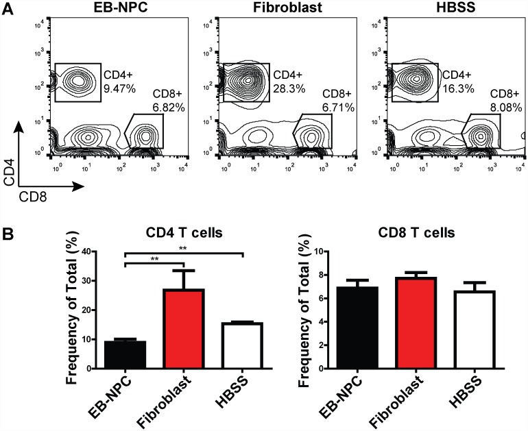
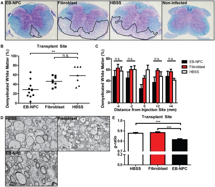
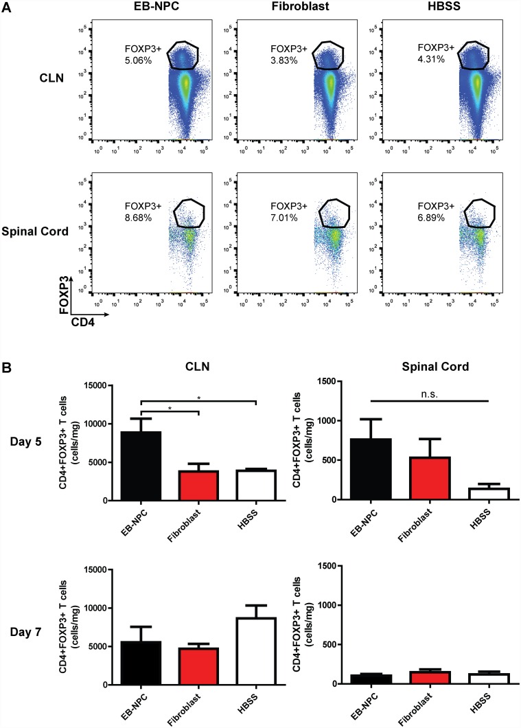
We have recently described sustained clinical recovery associated with dampened neuroinflammation and remyelination following transplantation of neural precursor cells (NPCs) derived from human embryonic stem cells (hESCs) in a viral model of the human demyelinating disease multiple sclerosis. The hNPCs used in that study were derived by a novel direct differentiation method (direct differentiation, DD-NPCs) that resulted in a unique gene expression pattern when compared to hNPCs derived by conventional methods. Since the therapeutic potential of human NPCs may differ greatly depending on the method of derivation and culture, we wanted to determine whether NPCs differentiated using conventional methods would be similarly effective in improving clinical outcome under neuroinflammatory demyelinating conditions. For the current study, we utilized hNPCs differentiated from a human induced pluripotent cell line via an embryoid body intermediate stage (EB-NPCs). Intraspinal transplantation of EB-NPCs into mice infected with the neurotropic JHM strain of mouse hepatitis virus (JHMV) resulted in decreased accumulation of CD4+ T cells in the central nervous system that was concomitant with reduced demyelination at the site of injection. Dampened neuroinflammation and remyelination was correlated with a transient increase in CD4+FOXP3+ regulatory T cells (Tregs) concentrated within the peripheral lymphatics. However, compared to our earlier study, pathological improvements were modest and did not result in significant clinical recovery. We conclude that the genetic signature of NPCs is critical to their effectiveness in this model of viral-induced neurologic disease. These comparisons will be useful for understanding what factors are critical for the sustained clinical improvement.


Conflict of interest statement

Competing Interests: The authors have declared that no competing interests exist.

Figures

Fig 1
Fig 1. Lentivirus-transduced human iPSCs differentiate to multipotent neural precursor cells.
(A) Representative micrographs of human iPSCs transduced with a lentiviral vector containing the Photinus pyralis luciferase gene and puromycin resistance cassette. Photon emission in response to D-luciferin could be detected in transduced puromycin-selected iPSCs (bottom) but not in non-transduced iPSCs (top) in vitro. (B) Representative FACS plots demonstrating CD184+/CD271-/CD44-/CD24+ gating scheme for isolation of NPCs from differentiated iPSCs. (C) Representative dot plots and quantification of NPC marker expression in FACS-sorted EB-NPCs following recovery and propagation (n = 3); data presented as average±SEM.
Fig 2
Fig 2. Human iPSC-derived NPCs are rapidly rejected following intraspinal transplantation in JHMV-infected mice.
(A) Timeline highlighting the relationship of T cell infiltration and demyelination in response to JHMV infection of the CNS as well as when cells are transplanted into the spinal cord and when animals are sacrificed to assess histology and immune cell infiltration into the CNS. (B) In vivo bioluminescence imaging revealed EB-NPCs could be detected in the spinal cords of transplanted animals as early as day 1 post-transplant (p.t.) and were undetectable by day 8 pt. (C) Representative brightfield images of coronal spinal cord sections from EB-NPC transplanted mice stained with SC121, a monoclonal antibody specific for human cytoplasm. Arrows indicate SC121+ regions. Human cells were detected in ventral white matter regions at day 1 p.t. but could be not be detected by day 8 p.t., confirming rejection of EB-NPCs. Scale bars = 100 μm.
Fig 3
Fig 3. CD4 T cell accumulation is reduced in EB-NPC-transplanted mice.
(A) Representative FACS plots demonstrating the frequency of CD4+ and CD8+ cells in the spinal cords of EB-NPC, human fibroblast, and HBSS-injected mice at day 21 pt. (B) Quantification of the frequency of CD4 and CD8 T cells. The frequency of CD4 T cells was significantly (p < 0.01) reduced in EB-NPC-transplanted animals compared to animals injected with human fibroblasts or HBSS. Data represents two independent experiments with n = 6 (EB-NPC), n = 4 (fibroblast), and n = 5 (HBSS) animals per group. Data presented as average+SEM and was analyzed using one-way ANOVA followed by Tukey’s multiple comparison test.
Fig 4
Fig 4. Focal remyelination in animals transplanted with EB-NPCs.
(A) Representative brightfield images of coronal spinal cord sections stained with luxol fast blue (LFB) and counter-stained with hemotoxylin and eosin (H&E). (B) Quantification of demyelination in the ventral white matter of EB-NPC, fibroblast, and HBSS injected mice revealed significantly (p < 0.01) reduced demyelination at the injection site in the spinal cords of EB-NPC-transplanted mice. (C) Quantification of demyelination in areas adjacent to the injection site revealed that reduced demyelination was not sustained along the rostrocaudal axis. (D) Representative electron micrographs of coronal spinal cord sections from HBSS, fibroblast, and EB-NPC-injected mice. (E) Analysis of the ratio of the axon diameter vs. total fiber diameter (g-ratio) confirmed enhanced remyelination at the transplant site of EB-NPC-injected mice compared to controls (p < 0.001). For (B) and (C), data represents two independent experiments with n = 11 (EB-NPC), n = 8 (Fibroblast), and n = 7 (HBSS) animals per group. For (D), ≥ 300 axons were measured per experimental group. All data is presented as average ± SEM and was analyzed using one-way ANOVA followed by Tukey’s multiple comparison test.
Fig 5
Fig 5. Regulatory T cells are increased in the draining cervical lymph nodes as a result of transient EB-NPC engraftment.
(A) Representative FACS plots of CD4+FOXP3+ cell analysis from the draining cervical lymph nodes (CLN; top) and spinal cords (bottom) of mice injected with EB-NPCs, fibroblasts, or HBSS. (B) Quantification of the number of CD4+FOXP3+ Tregs demonstrated a significant (p < 0.05) increase in the CLNs of EB-NPC transplanted mice compared to controls at day 5 p.t. (top). An increase in Tregs was not detected in the CLNs or spinal cords of EB-NPC transplanted mice by day 7 p.t. (bottom). Data represents two independent experiments. For day 5 analysis, n = 4 (EB-NPC), n = 6 (Fibroblast), and n = 4 (HBSS) animals per group. For day 7 analysis, n = 6 (EB-NPC), n = 7 (Fibroblast), and n = 5 (HBSS) animals per group. Data is presented as average ± SEM and was analyzed using one-way ANOVA followed by Tukey’s multiple comparison test.
Fig 6
Fig 6. Human iPSC-derived NPCs directly induce Treg conversion in vitro.
(A) Representative FACS plots of CD4+FOXP3+ Tregs from EB-NPC-T cell co-cultures. T cells were mixed with EB-NPCs at varying T cell-to-EB-NPC ratios and activated in the presence of anti-CD3/anti-CD28 beads for three days. (B) The frequency and number of FOXP3-expressing CD4+ T cells was significantly increased in the presence of EB-NPCs. (C) The observed increase in Tregs was correlated with a significant decrease in the number of conventional CD4+ T cells at a T cell-to-EB-NPC ratio of 1:4. All data was analyzed using students t-test and is presented as average ± SEM; *p < 0.05; **p < 0.01; ***p < 0.001; n = 4.
Fig 7
Fig 7. Regulatory T cells are necessary for EB-NPC-induced myelin sparing.
(A) Treatment of EB-NPC-transplanted mice with PC61.5, a rat monoclonal antibody raised against CD25, resulted in a significant reduction in the frequency of circulating CD4+FOXP3+ cells that was sustained to day 21 p.t. (B) Representative spinal cord sections stained with LFB and H&E. Outlined areas highlight demyelination. (C) Quantification of white matter damage revealed PC61.5-treated EB-NPC-transplanted mice did not have reduced demyelination when compared to non-treated EB-NPC-transplanted mice. For (A) and (C), data represents two independent experiments with n = 8 (EB-NPC alone), n = 9 (EB-NPC + IgG control), n = 9 (EB-NPC + PC61.5), and n = 8 (HBSS) animals per group. All data is presented as the average ± SEM and was analyzed using one-way ANOVA followed by Tukey’s multiple comparison test; *p < 0.05, **p < 0.01, ***p < 0.001.
Fig 8
Fig 8. EB-NPCs are significantly different than our previously differentiated NPCs.
EB-NPCs cluster away from both pluripotent stem cells and NPCs derived from both ESCs and iPSCs by our previously published method in both (A) Principal component analysis and (B) hierarchical clustering. (C) EB-NPCs show a lack of TGFβ2 transcript as shown by whole genome expression analysis.

Similar articles

Cited by

References

    1. Compston A, Coles A. Multiple sclerosis. Lancet. 2008;372(9648):1502–17. Epub 2008/10/31. 10.1016/S0140-6736(08)61620-7 . - DOI - PubMed
    1. Steinman L, Martin R, Bernard C, Conlon P, Oksenberg JR. Multiple sclerosis: deeper understanding of its pathogenesis reveals new targets for therapy. Annu Rev Neurosci. 2002;25:491–505. Epub 2002/06/08. 10.1146/annurev.neuro.25.112701.142913 . - DOI - PubMed
    1. Noseworthy JH, Lucchinetti C, Rodriguez M, Weinshenker BG. Multiple sclerosis. N Engl J Med. 2000;343(13):938–52. Epub 2000/09/28. 10.1056/NEJM200009283431307 . - DOI - PubMed
    1. Blurton-Jones M, Kitazawa M, Martinez-Coria H, Castello NA, Muller FJ, Loring JF, et al. Neural stem cells improve cognition via BDNF in a transgenic model of Alzheimer disease. Proc Natl Acad Sci U S A. 2009;106(32):13594–9. Epub 2009/07/28. 10.1073/pnas.0901402106 - DOI - PMC - PubMed
    1. McBride JL, Behrstock SP, Chen EY, Jakel RJ, Siegel I, Svendsen CN, et al. Human neural stem cell transplants improve motor function in a rat model of Huntington's disease. J Comp Neurol. 2004;475(2):211–9. Epub 2004/06/24. 10.1002/cne.20176 . - DOI - PubMed

MeSH terms