. 2016 Jun 16;11(6):e0156907.
doi: 10.1371/journal.pone.0156907. eCollection 2016.

BarTeL, a Genetically Versatile, Bioluminescent and Granule Neuron Precursor-Targeted Mouse Model for Medulloblastoma

Affiliations

BarTeL, a Genetically Versatile, Bioluminescent and Granule Neuron Precursor-Targeted Mouse Model for Medulloblastoma

Gregory M Shackleford et al. PLoS One. .

Abstract

Medulloblastomas are the most common malignant pediatric brain tumor and have been divided into four major molecular subgroups. Animal models that mimic the principal molecular aberrations of these subgroups will be important tools for preclinical studies and allow greater understanding of medulloblastoma biology. We report a new transgenic model of medulloblastoma that possesses a unique combination of desirable characteristics including, among others, the ability to incorporate multiple and variable genes of choice and to produce bioluminescent tumors from a limited number of somatic cells within a normal cellular environment. This model, termed BarTeL, utilizes a Barhl1 homeobox gene promoter to target expression of a bicistronic transgene encoding both the avian retroviral receptor TVA and an eGFP-Luciferase fusion protein to neonatal cerebellar granule neuron precursor (cGNP) cells, which are cells of origin for the sonic hedgehog (SHH) subgroup of human medulloblastomas. The Barhl1 promoter-driven transgene is expressed strongly in mammalian cGNPs and weakly or not at all in mature granule neurons. We efficiently induced bioluminescent medulloblastomas expressing eGFP-luciferase in BarTeL mice by infection of a limited number of somatic cGNPs with avian retroviral vectors encoding the active N-terminal fragment of SHH and a stabilized MYCN mutant. Detection and quantification of the increasing bioluminescence of growing tumors in young BarTeL mice was facilitated by the declining bioluminescence of their uninfected maturing cGNPs. Inclusion of eGFP in the transgene allowed enriched sorting of cGNPs from neonatal cerebella. Use of a single bicistronic avian vector simultaneously expressing both Shh and Mycn oncogenes increased the medulloblastoma incidence and aggressiveness compared to mixed virus infections. Bioluminescent tumors could also be produced by ex vivo transduction of neonatal BarTeL cerebellar cells by avian retroviruses and subsequent implantation into nontransgenic cerebella. Thus, BarTeL mice provide a versatile model with opportunities for use in medulloblastoma biology and therapeutics.


Conflict of interest statement

Competing Interests: The authors have declared that no competing interests exist.

Figures

Fig 1
Fig 1. Barhl1 is expressed in cerebellum of neonatal mice and medulloblastoma cell lines, and an isolated Barhl1 promoter is active in medulloblastoma cell lines.
(A) RT-PCR analysis of Barhl1 expression in mouse cerebellum and non-cerebellar brain tissue at postnatal days 3, 7, 11 and 24. The PCR cycle number used was limited for Barhl1 (32 cycles) and Gapdh (23 cycles) in order to enable distinction of RNA levels in cerebellum samples; high cycle number (40 cycles) showed only a faint Barhl1 product in non-cerebellum lanes. The last lane contains 100-bp markers. PCR Primers are listed in S2 Table. (B) RT-PCR analysis of the expression of BARHL1, NESTIN and ATOH1 in human medulloblastoma, glioblastoma and atypical teratoid/rhabdoid tumor cell lines. All PCRs were 40 cycles except GAPDH (23 cycles). PCR Primers are listed in S2 Table. (C) Expression of BARHL1 in the four subgroups of human medulloblastoma. BARHL1 gene expression data were obtained from the R2 genomic analysis and visualization platform and plotted according to subgroup. The database used was “Tumor Medulloblastoma (core transcript)–Northcott– 103 –rma_sketch–huex10t”. (D) Testing the Barhl1 promoter in a luciferase reporter assay for activity in medulloblastoma cell lines. The Barhl1-luciferase reporter plasmid was co-transfected with a constitutive Renilla luciferase control plasmid into the cell lines shown. Data were corrected for transfection efficiency. The first lane contains 100-bp markers.
Fig 2
Fig 2. The BarTeL transgene is specifically expressed in EGL of neonatal mice.
(A) Structure of the BarTeL transgene and RCAS retroviruses. In BarTeL, the mouse Barhl1 promoter drives expression of a quail Tva cDNA. An IRES allows co-expression of an eGFP-luciferase fusion protein. The RCAS replication competent retroviral vectors express the inserted oncogenes via a splice acceptor located immediately upstream of the cloning site.(B) Bioluminescence imaging shows expression of the BarTeL transgene in the cerebellar region of three transgenic lines. Pups from lines 1–2, 2–1 and 6–4 were injected with luciferin and examined by optical imaging for bioluminescence. Age at imaging is indicated under the panels. (C) Histology and immunohistochemistry for luciferase transgene protein in brains of BarTeL mouse pups. H&E and successive magnifications (boxed, clockwise) of anti-luciferase staining of sagittal sections of transgenic mouse pup cerebella are shown. (D) eGFP expression in BarTeL cerebellar cells. Dissociated cerebella from BarTeL and nontransgenic mouse pups were analyzed by flow cytometry to demonstrate eGFP expression and the ability to sort eGFP-positive cells, indicative of BarTeL transgene expression.
Fig 3
Fig 3. ShhN and MycnT50A,S54A cooperatively induce bioluminescent medulloblastomas in vivo in BarTeL mice.
(A) BarTeL mice were injected with a mixture of chicken DF-1 cells producing RCAS-ShhN and RCAS-MycnT50A,S54A viruses and imaged for bioluminescence starting at 14 days post-injection. Shown are images at two week intervals of four mice that developed bioluminescent tumors (left) compared to three mice without tumor (right). (B) Time course of bioluminescent tumor formation in transgenic mice. A cohort of 11 transgenic mice, 10 of which were infected as in (A), were imaged weekly for 12 weeks. At each time point, net ΔLog10 flux values plotted for each mouse were determined by subtracting the mean of the log values of flux of non-tumor-bearing transgenic mice from the log of the flux of each mouse. A graph of unprocessed measurements before conversion to log values is provided separately (S2 Fig) to show the loss of bioluminescence by cGNPs during postnatal cerebellar development and the concomitant growth of tumor bioluminescence in those mice that developed medulloblastomas. (C) Medulloblastoma in whole BarTeL brain and immunohistochemistry for SHHN and MYCN. Gross appearance of a medulloblastoma in a formalin-fixed brain (second panel) compared to a normal brain (first panel) from a littermate. Staining with an anti-SHH antibody showed that only a minority of medulloblastoma cells produced SHHN protein (fourth panel). Purkinje cells in control normal (NL) adult cerebellum also stain positively with anti-SHH antibody (third panel). An anti-c-MYC antibody identified the MYC-tagged MYCNT50A,S54A protein in most cells of the same tumor (fifth panel). The magnified panels above the SHH and MYCN panels show the hypercellularity of these tumors. Bar, 10 μm. (D) Histology and immunohistochemistry of a representative medulloblastoma induced by ShhN and MycnT50A,S54A viruses. Shown are an H&E-stained tumor section (left panel) and tumor sections stained with antibodies to PGP9.5 (ubiquitin carboxyl-terminal esterase L1), Synaptophysin, Ki67 and GFAP. Bar, 10 μm.
Fig 4
Fig 4. Neonatal BarTeL cGNP cells infected ex vivo with a mix of RCAS-ShhN and RCAS-MycnT50A,S54A viruses efficiently develop into bioluminescent medulloblastomas when orthotopically injected into nontransgenic cerebella.
(A) PCR assay to detect the specific infection of transgenic cGNPs in culture. Cerebella from transgenic (Tg) and non-transgenic wild type (WT) mice were dissociated, put into culture, infected with RCAS-ShhN and RCAS-MycnT50A,S54A viruses and then used for preparation of genomic DNA as described in Materials and Methods. PCRs of these genomic DNAs were performed to test for presence of RCAS-ShhN (upper panels) or RCAS-MycnT50A,S54A (lower panels) proviral DNAs. Positive control template (+) was DNA from a ShhN and MycnT50A,S54A-induced mouse medulloblastoma, and negative control template (–) was DNA from an uninfected transgenic cerebellum. Control primers that amplify an intron region of a single-copy gene (Fgfr2) were included in all PCRs. The third lane contains 100-bp markers. Thirty-five PCR cycles were used. PCR Primers are listed in S2 Table. (B) Bioluminescent tumors induced by transplanting ex vivo infected transgenic cerebellar cells into non-transgenic cerebella. Dissociated cerebellar cells from transgenic (Tg) or non-transgenic (WT) neonatal mice were exposed to RCAS-ShhN and RCAS-MycnT50A,S54A viruses in culture, harvested, injected into the cerebella of non-transgenic neonatal B6D2F1 mice and imaged at the times shown. (C) Time course of bioluminescent tumor formation in recipient mice orthotopically injected with ex vivo-infected transgenic cerebellar cells. Bioluminescence measurements of the mice shown in (B) were plotted over time. Solid lines (blue) denote mice receiving transgenic cerebellar cells; dotted line (black, empty circles) denotes mouse receiving non-transgenic cells.
Fig 5
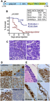
Fig 5. Bicistronic RCAS virus expressing both MycnT58A and ShhN increases tumor incidence and lethality.
(A) Map of the bicistronic RCAS vector. Mycn* denotes MycnT58A. Not to scale. (B) Kaplan-Meier curves of time to tumor-related euthanasia of mice infected with the RCASBP(A)ΔF1’-MycnT58A/ShhN bicistronic virus (maroon) compared to a mixture of the RCASBP(A)ΔF1’-MycnT58A and RCASBP(A)ΔF1’-ShhN single-gene viruses (blue). The inset table shows the incidence of medulloblastomas in BarTeL mice infected with the bicistronic virus (ShhN/Mycn), the separate single-gene viruses and a mixture of the two single-gene viruses (ShhN + Mycn) derived from the bicistronic plasmid. Mice were euthanized upon development of tumor-related symptoms. (C) H&E stain of a BarTeL bicistronic medulloblastoma. Typical field from an H&E stain of a medulloblastoma arising in the cerebellum of a BarTeL mouse that was injected with the ShhN/Mycn-containing bicistronic virus. Right panel is a magnified view. Bar, 10 μm. (D) Immunohistochemical staining of a representative medulloblastoma (ShhN/Mycn bicistronic virus) with antibodies to the indicated proteins. Luciferase staining demonstrates the border between normal cerebellum (nl, negative) and the tumor (MB; positive). MYCN expression is detected by staining with antibody to the human MYC epitope tag and shows that almost all cells in the tumor express nuclear MycnT58A. Ki67 staining shown is at the border of the tumor, with the upper left area being adjacent normal brain. SHH staining is shown in medulloblastoma (MB) compared to the normal (NL) cerebellum, the latter of which demonstrates SHH expression in three Purkinje cells at the border between the molecular layer (upper left in image) and the IGL. Negative control of medulloblastoma was stained with nonspecific IgG primary antibody. Bar, 10 μm. (E) Leptomeningeal dissemination and spread in brains from bicistronic virus-infected mice. Shown are an H&E stain (upper left; note the overlying meninges) and staining with antibodies to luciferase (lower left) and the MYC epitope tag of MYCNT58A (lower right) of areas in the same leptomeningeal dissemination over the hemisphere. In a different mouse, the upper right panel demonstrates leptomeningeal spread over normal cerebellum, which is contiguous to the main tumor mass a few millimeters away, outside the right lower area of the panel. Bar, 10 μm.
Fig 6
Fig 6. Molecular subgrouping analysis of BarTeL ShhN/MycnT58A medulloblastomas.
Scatter plot of the first two principle components (PC) of human medulloblastoma samples (GSE4036) and BarTeL Shh/MycnT58A (bicistronic virus-induced) medulloblastomas. Principle component analysis used a subset of highly variable genes (n = 631) among human medulloblastoma. The expression data from human genes and their mouse orthologs were normalized against the corresponding species-specific normal cerebella.

Similar articles

Cited by

References

    1. Packer RJ. Childhood brain tumors: accomplishments and ongoing challenges. J Child Neurol. 2008;23(10):1122–7. doi: 23/10/1122 [pii] 10.1177/0883073808320758 - DOI - PMC - PubMed
    1. Rutkowski S, von Hoff K, Emser A, Zwiener I, Pietsch T, Figarella-Branger D, et al. Survival and prognostic factors of early childhood medulloblastoma: an international meta-analysis. J Clin Oncol. 2010;28(33):4961–8. doi: JCO.2010.30.2299 [pii] 10.1200/JCO.2010.30.2299 . - DOI - PubMed
    1. Thompson MC, Fuller C, Hogg TL, Dalton J, Finkelstein D, Lau CC, et al. Genomics identifies medulloblastoma subgroups that are enriched for specific genetic alterations. J Clin Oncol. 2006;24(12):1924–31. 10.1200/JCO.2005.04.4974 . - DOI - PubMed
    1. Kool M, Koster J, Bunt J, Hasselt NE, Lakeman A, van Sluis P, et al. Integrated genomics identifies five medulloblastoma subtypes with distinct genetic profiles, pathway signatures and clinicopathological features. PLoS ONE. 2008;3(8):e3088 10.1371/journal.pone.0003088 . - DOI - PMC - PubMed
    1. Cho YJ, Tsherniak A, Tamayo P, Santagata S, Ligon A, Greulich H, et al. Integrative genomic analysis of medulloblastoma identifies a molecular subgroup that drives poor clinical outcome. J Clin Oncol. 2011;29(11):1424–30. 10.1200/JCO.2010.28.5148 - DOI - PMC - PubMed

MeSH terms

LinkOut - more resources