

Myeloid sarcoma of the Gingiva with myelodysplastic syndrome: A Case Report
- PMID: 27310987
- PMCID: PMC4998473
- DOI: 10.1097/MD.0000000000003897
Myeloid sarcoma of the Gingiva with myelodysplastic syndrome: A Case Report
Erratum in
-
Erratum: Medicine, Volume 95, Issue 24: Erratum.Medicine (Baltimore). 2016 Aug 7;95(31):e5074. doi: 10.1097/01.md.0000490009.39850.74. eCollection 2016 Aug. Medicine (Baltimore). 2016. PMID: 31265618 Free PMC article.
Abstract
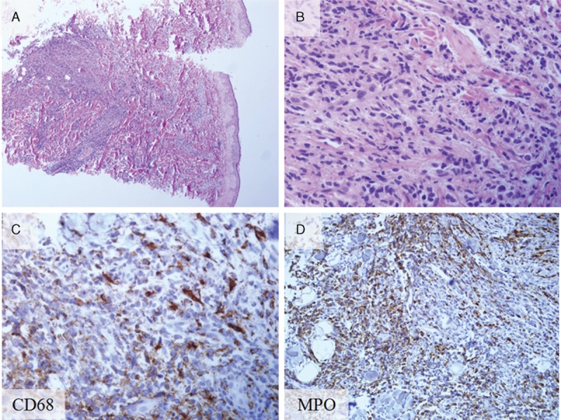
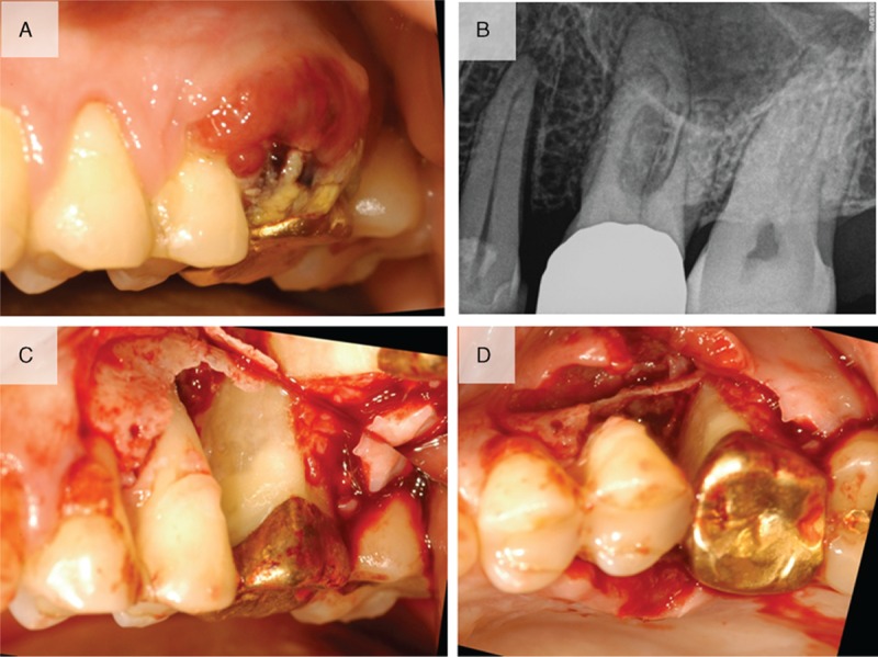
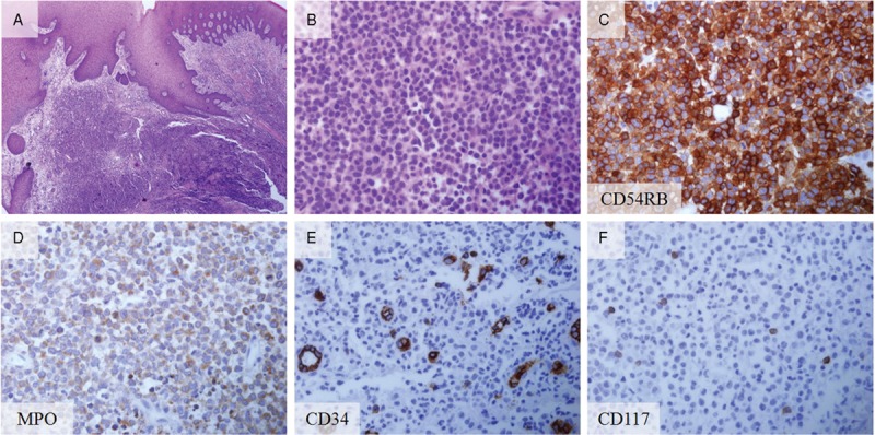
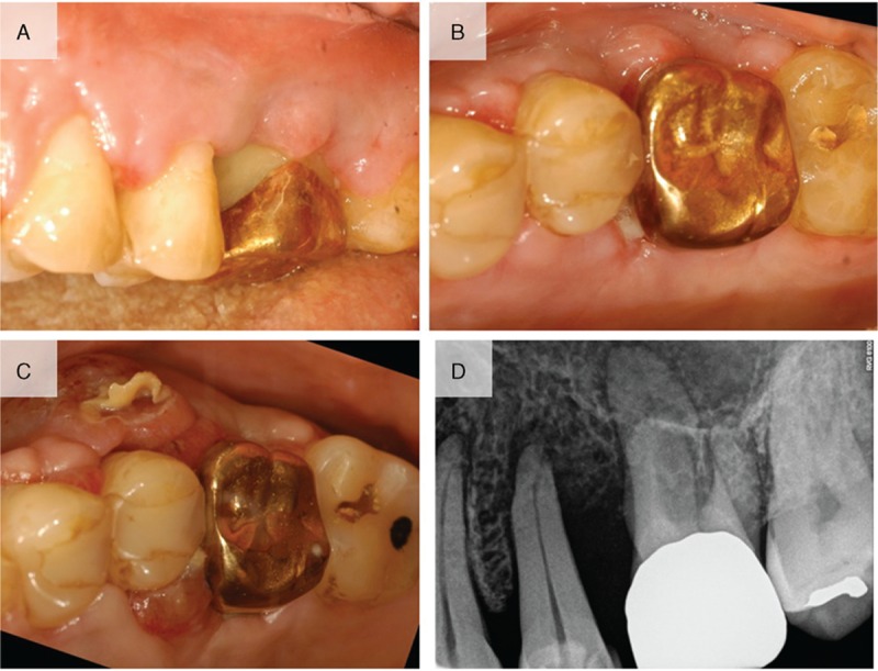
The purpose of this report is to present a case of myeloid sarcoma of the gingiva with myelodysplastic syndrome.A 52-year-old male diagnosed with myelodysplastic syndrome with skin lesions presented with gingival swelling and gingival redness involving the maxillary left second premolar and the maxillary left first molar. The patient was referred from the Department of Hematology for a biopsy of the lesion. Full-thickness flaps were elevated and inflamed, and neoplastic soft tissue was removed from a lesion and the samples sent for histopathologic analysis.Histopathologic results showed leukemic cell infiltration beneath the oral epithelium, and the specimen was positive for the leukocyte marker. The diagnosis was myeloid sarcoma. Uneventful healing was observed at 2-week follow-up, but relapse of the lesions with the hyperplastic and neoplastic tissue was noted at 4-week follow-up. Further follow-up or treatment could not be performed because the patient did not visit at the next follow-up.In conclusion, myeloid sarcoma should be a diagnosis option for gingival growth because it can involve intraoral lesion. In this report, a biopsy was performed due to referral considering the patient's medical history. Although myeloid sarcoma in the oral cavity is extremely rare, a small biopsy and consultation with a hematologist may be beneficial for patients and may provide a differential diagnosis.
Conflict of interest statement
The authors have no conflicts of interest to disclose.
Figures
References
-
- Antmen B, Haytac MC, Sasmaz I, et al. Granulocytic sarcoma of gingiva: an unusual case with aleukemic presentation. J Periodontol 2003;74:1514–9. - PubMed
-
- Liu PI, Ishimaru T, McGregor DH, et al. Autopsy study of granulocytic sarcoma (chloroma) in patients with myelogenous leukemia, Hiroshima-Nagasaki 1949–1969. Cancer 1973;31:948–55. - PubMed
-
- Falini B, Lenze D, Hasserjian R, et al. Cytoplasmic mutated nucleophosmin (NPM) defines the molecular status of a significant fraction of myeloid sarcomas. Leukemia 2007;21:1566–70. - PubMed
-
- Pileri SA, Ascani S, Cox MC, et al. Myeloid sarcoma: clinico-pathologic, phenotypic and cytogenetic analysis of 92 adult patients. Leukemia 2007;21:340–50. - PubMed
Publication types
MeSH terms
LinkOut - more resources
Full Text Sources
Other Literature Sources
Medical
