

Endoscopic removal of bullets from the cranial ridge junction region via transoral and transnasal approaches: Two case reports and review of literature
- PMID: 27310999
- PMCID: PMC4998485
- DOI: 10.1097/MD.0000000000003918
Endoscopic removal of bullets from the cranial ridge junction region via transoral and transnasal approaches: Two case reports and review of literature
Erratum in
-
Erratum: Medicine, Volume 95, Issue 24: Erratum.Medicine (Baltimore). 2016 Aug 7;95(31):e5074. doi: 10.1097/01.md.0000490009.39850.74. eCollection 2016 Aug. Medicine (Baltimore). 2016. PMID: 31265618 Free PMC article.
Abstract
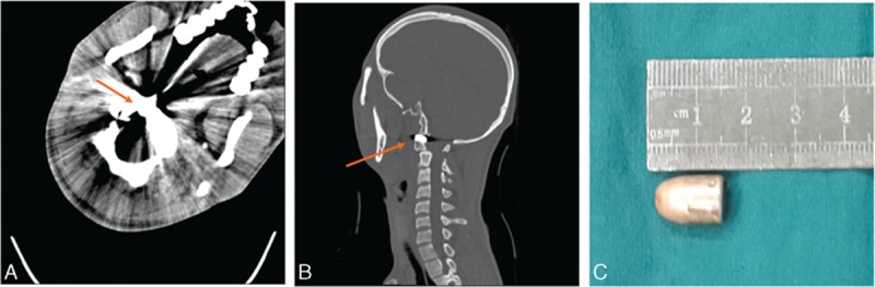
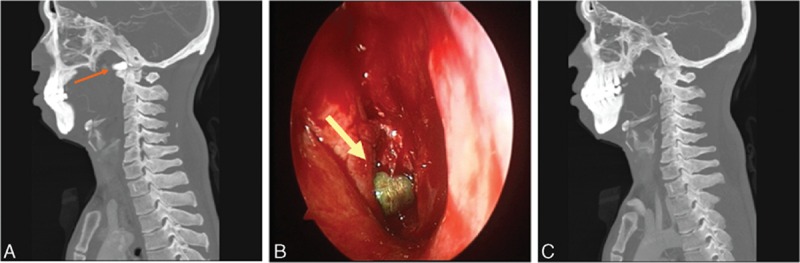
Endoscopes in otolaryngology may facilitate accessing the lumens and sites such as upper cervical spine with minimally invasive surgical exposure. Here, we present 2 interesting cases of youth who underwent endoscopic removal of bullets in the cranial ridge junction region.The first case was a 20-year-old young man who underwent a gunshot in the face. A CT scan showed that a metallic foreign body located inside the right lateral body of Atlas that presented a comminuted fracture. The second case a 36-year-old man who also underwent a gunshot in the face. CT scan showed a foreign body lodged in the soft tissues before the right anterior arch of Atlas cone (C1) that presented a fracture. The bullets in these 2 patients were removed under the endoscopes with minimal damage, respectively. The patients were discharged without neck activity obstacle.The advantage of endoscopic technique is obvious because limited visualization does not damage surrounding tissues, thus decreasing surgical complications. This was an interesting experience of surgical operation in this region.
Conflict of interest statement
The authors have no funding and conflicts of interest to disclose.
Figures
References
Publication types
MeSH terms
LinkOut - more resources
Full Text Sources
Other Literature Sources
Medical
