

Scabies: Advances in Noninvasive Diagnosis
- PMID: 27311065
- PMCID: PMC4911127
- DOI: 10.1371/journal.pntd.0004691
Scabies: Advances in Noninvasive Diagnosis
Abstract
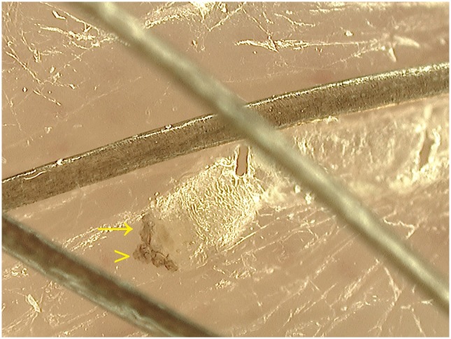
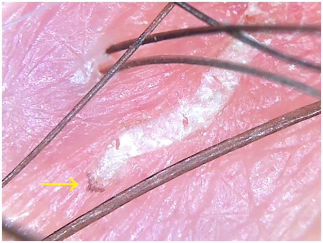
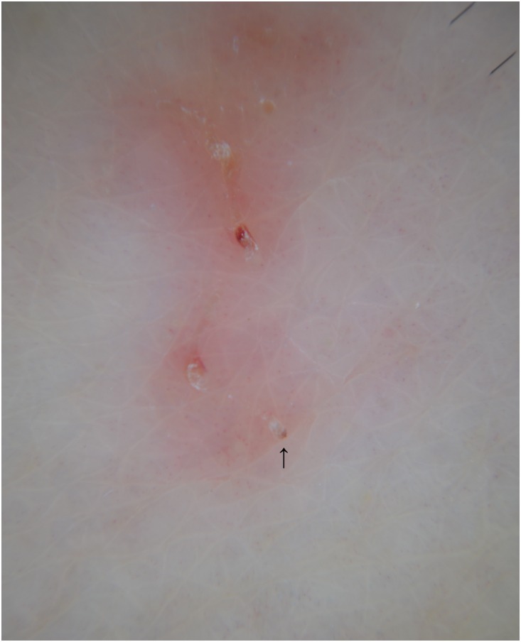
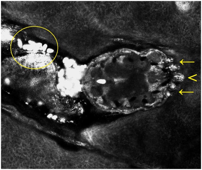
Scabies is a common, highly contagious skin parasitosis caused by Sarcoptes scabiei var. hominis. Early identification and prompt treatment of infested subjects is essential, as missed diagnosis may result in outbreaks, considerable morbidity, and significantly increased economic burden. The standard diagnostic technique consists of mites' identification by microscopic examination of scales obtained by skin scraping. This is a time-consuming and risk-associated procedure that is also not suitable to a busy practice. In recent years, some advanced and noninvasive techniques such as videodermatoscopy, dermatoscopy, reflectance confocal microscopy, and optical coherence tomography have demonstrated improved efficacy in the diagnosis of scabies. Their advantages include rapid, noninvasive mass screening and post-therapeutic follow-up, with no physical risk. A greater knowledge of these techniques among general practitioners and other specialists involved in the intake care of overcrowded populations vulnerable to scabies infestations is now viewed as urgent and important in the management of outbreaks, as well as in consideration of the recent growing inflow of migrants in Europe from North Africa.
Conflict of interest statement
The authors have declared that no competing interests exist.
Figures
References
-
- Hengge UR, Currie BJ, Jager G, Lupi O, Schwartz RA. Scabies: a ubiquitous neglected skin disease. Lancet Infect Dis 2006;6:769–779. - PubMed
-
- Heukelbach J, Feldmeier H. Scabies. Lancet 2006;367:1767–1774. - PubMed
-
- Feldmeier H, Jackson A, Ariza L, Calheiros CM, Soares Vde L, Oliveira FA, et al. The epidemiology of scabies in an impoverished community in rural Brazil: presence and severity of disease are associated with poor living conditions and illiteracy. J Am Acad Dermatol 2009;60:436–443. 10.1016/j.jaad.2008.11.005 - DOI - PubMed
Publication types
MeSH terms
LinkOut - more resources
Full Text Sources
Other Literature Sources
Medical
