Review
. 2016 Aug:44:33-41.
doi: 10.1016/j.dnarep.2016.05.004. Epub 2016 May 13.

Involvement of translesion synthesis DNA polymerases in DNA interstrand crosslink repair

Affiliations
Review

Involvement of translesion synthesis DNA polymerases in DNA interstrand crosslink repair

Upasana Roy et al. DNA Repair (Amst). 2016 Aug.

Abstract

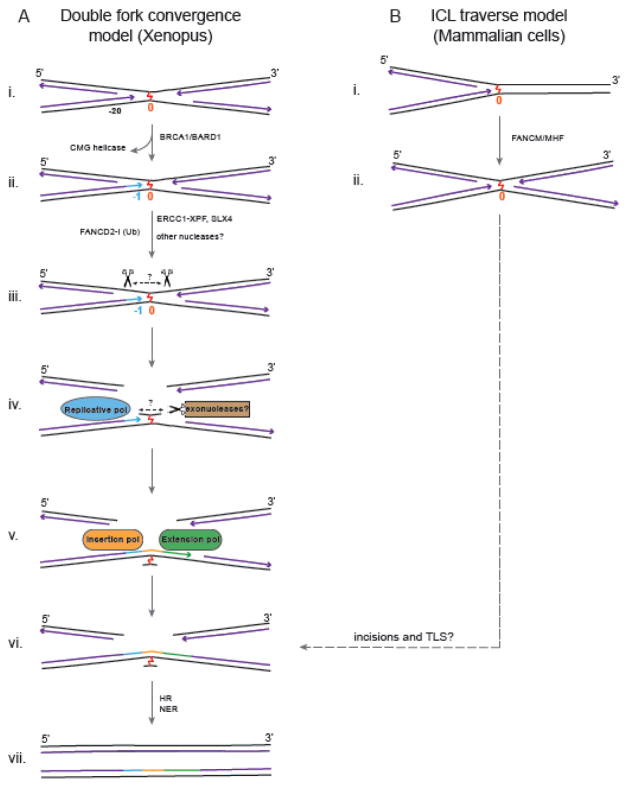
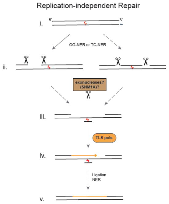
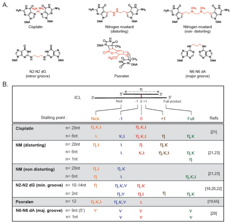
DNA interstrand crosslinks (ICLs) covalently join the two strands of a DNA duplex and block essential processes such as DNA replication and transcription. Several important anti-tumor drugs such as cisplatin and nitrogen mustards exert their cytotoxicity by forming ICLs. However, multiple complex pathways repair ICLs and these are thought to contribute to the development of resistance towards ICL-inducing agents. While the understanding of many aspects of ICL repair is still rudimentary, studies in recent years have provided significant insights into the pathways of ICL repair. In this perspective we review the recent advances made in elucidating the mechanisms of ICL repair with a focus on the role of TLS polymerases. We describe the emerging models for how these enzymes contribute to and are regulated in ICL repair, discuss the key open questions and examine the implications for this pathway in anti-cancer therapy.

Keywords: Cisplatin; DNA polymerases; Inter-strand crosslink repair; Nitrogen mustard; Translesion synthesis.


Figures

Fig. 1
Fig. 1. Models for Replication-Dependent ICL Repair Pathways
A: Double Fork Convergence Model. (i) Two replication forks converge on an ICL and stall 20–40 nt away from it. (ii) Removal of the CMG helicase by BRCA1 allows one of the leading strands to approach within 1 nt of the ICL (−1). (iii) Activation of the FA pathway leads to ubiquitylation of FANCD2-I, which is required for unhooking of the ICL by SLX4/ERCC1-XPF and possibly other nucleases. The position of these incisions have not been determined, and the amount of duplex surrounding the ICL is unknown. (iv) The unhooked ICL could then be further processed by exonucleases to trim the duplex around the ICL, making it more amenable to bypass by DNA polymerases. (v, vi) An insertion polymerase inserts nucleotide(s) opposite the ICL and an extension polymerase extends the insertion product further. (vi) Ligation to downstream Okazaki fragments restores one daughter duplex, and (vii) is used to restore the other duplex by HR. The ICL remnant on one strand is likely removed by NER to complete the repair of both daughter duplexes. B: ICL traverse model. (i) A single fork collides with the ICL, and (ii) in a FANCM/MHF dependent manner ‘traverses’ the ICL to continue replication on the other side of the crosslink without unhooking it. The later steps of this pathway are not known, but could involve incisions and TLS for post-replicative repair.
Fig. 2
Fig. 2. Model for Replication-Independent ICL Repair
(i) Global genome NER (GG-NER) as well as transcription coupled NER (TC-NER) proteins are involved in replication independent repair of ICLs. (ii) Although dual incisions 5′ to the ICL have been observed, unhooking incisions on either side of the ICL may also occur. (iii) These intermediates may be further processed by exonucleases like SNM1A to facilitate translesion synthesis. (iv) TLS polymerases carry out gap filling and (v) the ICL remnant is likely removed by NER. Solid arrows indicate details obtained from experimental observations and dashed arrows indicate indirect evidence and/or speculation.
Fig. 3
Fig. 3. Biochemical Activity of TLS Polymerases on ICL Substrates
A: Chemical Structures of ICLs. The crosslinks between bases are highlighted in red. B: Primer Extension Activity of TLS Polymerases η,κ,ν and ι across diverse ICL substrates with varying amount of duplex (n) surrounding the crosslink. For polymerases that were tested with the substrates shown the main stalling points at the start of the duplex (nick, orange), 1 nt before the ICL (−1, blue), at the ICL (0, red), 1 nt after the ICL (+1, brown) as well as complete extension to full product (Full, green) are indicated for each ICL substrate. Although conditions used to generate the data listed in this figure varied greatly, ICLs embedded in a longer duplex were not bypassed efficiently by TLS polymerases.

References

    1. Clauson C, Schärer OD, Niedernhofer L. Advances in understanding the complex mechanisms of DNA interstrand cross-link repair. Cold Spring Harb Perspect Biol. 2013;5:a012732. - PMC - PubMed
    1. Kelland L. The resurgence of platinum-based cancer chemotherapy. Nat Rev Cancer. 2007;7:573–584. - PubMed
    1. Deans AJ, West SC. DNA interstrand crosslink repair and cancer. Nat Rev Cancer. 2011;11:467–480. - PMC - PubMed
    1. Kottemann MC, Smogorzewska A. Fanconi anaemia and the repair of Watson and Crick DNA crosslinks. Nature. 2013;493:356–363. - PMC - PubMed
    1. Räschle M, Knipscheer P, Enoiu M, Angelov T, Sun J, Griffith JD, Ellenberger TE, Schärer OD, Walter JC. Mechanism of replication-coupled DNA interstrand crosslink repair. Cell. 2008;134:969–980. - PMC - PubMed

Publication types

MeSH terms

LinkOut - more resources