. 2016 Oct;10(8):1221-31.
doi: 10.1016/j.molonc.2016.05.009. Epub 2016 Jun 7.

Droplet digital PCR of circulating tumor cells from colorectal cancer patients can predict KRAS mutations before surgery

Affiliations

Droplet digital PCR of circulating tumor cells from colorectal cancer patients can predict KRAS mutations before surgery

Jérôme Alexandre Denis et al. Mol Oncol. 2016 Oct.

Abstract

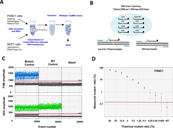
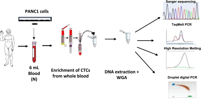
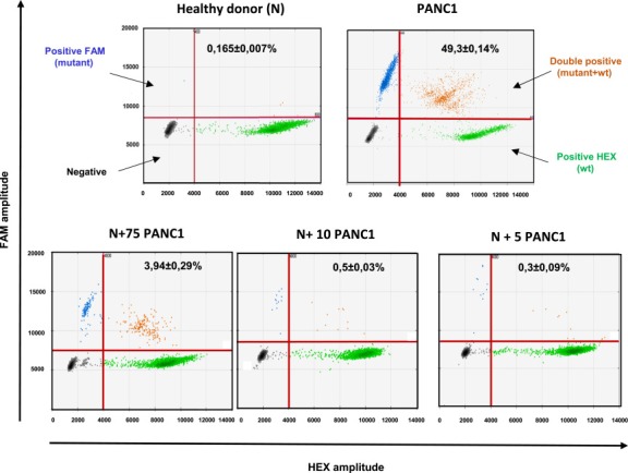
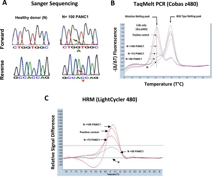
In colorectal cancer (CRC), KRAS mutations are a strong negative predictor for treatment with the EGFR-targeted antibodies cetuximab and panitumumab. Since it can be difficult to obtain appropriate tumor tissues for KRAS genotyping, alternative methods are required. Circulating tumor cells (CTCs) are believed to be representative of the tumor in real time. In this study we explored the capacity of a size-based device for capturing CTCs coupled with a multiplex KRAS screening assay using droplet digital PCR (ddPCR). We showed that it is possible to detect a mutant ratio of 0.05% and less than one KRAS mutant cell per mL total blood with ddPCR compared to about 0.5% and 50-75 cells for TaqMeltPCR and HRM. Next, CTCs were isolated from the blood of 35 patients with CRC at various stage of the disease. KRAS genotyping was successful for 86% (30/35) of samples with a KRAS codon 12/13 mutant ratio of 57% (17/30). In contrast, only one patient was identified as KRAS mutant when size-based isolation was combined with HRM or TaqMeltPCR. KRAS status was then determined for the 26 available formalin-fixed paraffin-embedded tumors using standard procedures. The concordance between the CTCs and the corresponding tumor tissues was 77% with a sensitivity of 83%. Taken together, the data presented here suggest that is feasible to detect KRAS mutations in CTCs from blood samples of CRC patients which are predictive for those found in the tumor. The minimal invasive nature of this procedure in combination with the high sensitivity of ddPCR might provide in the future an opportunity to monitor patients throughout the course of disease on multiple levels including early detection, prognosis, treatment and relapse as well as to obtain mechanistic insight with respect to tumor invasion and metastasis.

Keywords: Circulating tumor cells; Colorectal cancer; Digital PCR; KRAS; Liquid biopsy.


Figures

Figure 1
Figure 1
Sensitivity of the KRAS multiplex assay by droplet digital PCR. A. Protocol. DNA was extracted from 100 PANC1 cells bearing a heterozygous mutation c.35G > A in KRAS codon 12 (Gly12Asp) and 100 MCF7 cell lines (wild‐type for KRAS codon 12–13). After whole genome amplification, DNA from the two cell lines was mixed and droplet digital PCR was performed using a multiplex KRAS assay. B. KRAS screening assay. This TaqMan assay is designed to detect the seven most frequent mutations in codon 12 and codon 13 of KRAS. C. Experimental validation. 1D‐Dot plot. The blue histogram indicates the number of droplets considered as positive for mutant KRAS according to the fluorescence threshold. The green histogram corresponds to the number of wild‐type droplets. Mutant control, DNA containing a 1:1 mix of mutant and wildtype KRAS. WT control, DNA containing only KRAS wild‐type. Blank, water. D. Mutant ratio threshold determination (mutant copies/total copies in %) according to theoretical mutant ratio obtained by serial dilution. The red line indicates the detection threshold.
Figure 2
Figure 2
Validation of the protocol for KRAS testing. Whole blood was spiked with PANC1 cells harboring a KRAS mutation in codon 12–13 followed by size‐based enrichment and multiplex KRAS analysis using droplet digital PCR. N, “Normal” blood from healthy donors. WGA, Whole Genome Amplification.
Figure 3
Figure 3
Detection threshold of KRAS from whole blood using droplet digital PCR. 2D‐Dot plot. Fluorescence results are plotted as two‐dimensional dot plots (similar to the depiction of flow cytometry data). The region of these plots can be sequentially separated based on the fluorescence intensity of each droplet. Grey dots correspond to empty droplets. Green dots correspond to droplets containing wild‐type copies of KRAS on codon 12–13. Blue dots correspond to droplets containing at least one codon 12–13 KRAS mutation. Orange dots correspond to droplets containing at least one KRAS wild type copy and one mutant copy. These droplets are considered for the analysis. Abundance ratio (KRAS mutant copies/total copies) is indicated for each dot plots. Each analysis was performed at least in triplicate. N, blood from “normal” healthy donors with no spiking of mutant KRAS cells. PANC1, blood from “normal” healthy donors spiked with PANC1 cells bearing the heterozygous c.35G > A mutation in KRAS codon 12 (Gly12Asp).
Figure 4
Figure 4
Comparison of the sensitivity for detection of KRAS mutant cells in whole blood. A. Sanger sequencing. Considering the weak signal, the detection limit is close to 100 cells. As control, no signal was detected in blood from healthy donors or in blood spiked with 50 PANC1 cells. B. TaqMeltPCR: The presence of a mutation melting peak at the correct annealing temperature indicate the presence of KRAS mutations. The sample of healthy donor blood spiked with 50 PANC1 cells is considered at the detection limit when applying validation guidelines. When the assay is performed in “CIVD mode” recommended for clinical applications, the detection limit is 100 cells. C, High Melting Resolution (HRM). The detection limit (red line) is between 75 and 50 cells.

References

    1. Alix-Panabieres, C. , Pantel, K. , 2014. Technologies for detection of circulating tumor cells: facts and vision. Lab Chip 14, 57–62. - PubMed
    1. Amado, R.G. , Wolf, M. , Peeters, M. , Van Cutsem, E. , Siena, S. , Freeman, D.J. , Juan, T. , Sikorski, R. , Suggs, S. , Radinsky, R. , Patterson, S.D. , Chang, D.D. , 2008. Wild-type KRAS is required for panitumumab efficacy in patients with metastatic colorectal cancer. J. Clin. Oncol. 26, 1626–1634. - PubMed
    1. Andree, K.C. , van Dalum, G. , Terstappen, L.W. , 2016. Challenges in circulating tumor cell detection by the CellSearch system. Mol. Oncol. 10, (3) 395–407. - PMC - PubMed
    1. Barriere, G. , Fici, P. , Gallerani, G. , Fabbri, F. , Zoli, W. , Rigaud, M. , 2014. Circulating tumor cells and epithelial, mesenchymal and stemness markers: characterization of cell subpopulations. Ann. Transl Med. 2, 109 - PMC - PubMed
    1. Barriere, G. , Tartary, M. , Rigaud, M. , 2014. Epithelial mesenchymal transition: a new insight into the detection of circulating tumor cells. ISRN Oncol. 2012, 382010 - PMC - PubMed

Publication types

Substances