. 2016 Jun 20;43(6):393-404.
doi: 10.1016/j.jgg.2016.03.008. Epub 2016 Apr 1.

Filamentation of Metabolic Enzymes in Saccharomyces cerevisiae

Affiliations

Filamentation of Metabolic Enzymes in Saccharomyces cerevisiae

Qing-Ji Shen et al. J Genet Genomics. .

Abstract

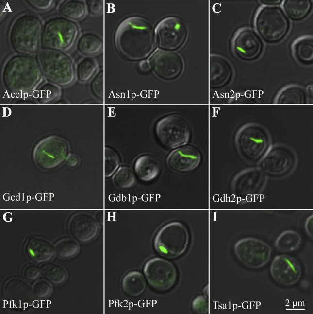
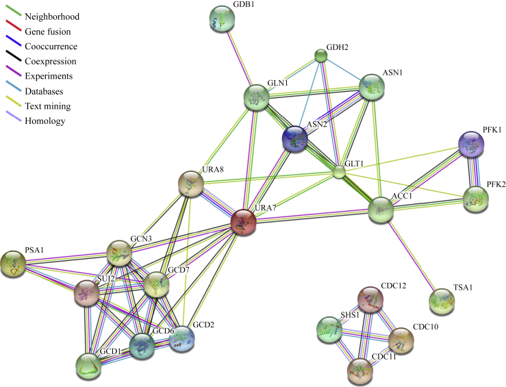
Compartmentation via filamentation has recently emerged as a novel mechanism for metabolic regulation. In order to identify filament-forming metabolic enzymes systematically, we performed a genome-wide screening of all strains available from an open reading frame-GFP collection in Saccharomyces cerevisiae. We discovered nine novel filament-forming proteins and also confirmed those identified previously. From the 4159 strains, we found 23 proteins, mostly metabolic enzymes, which are capable of forming filaments in vivo. In silico protein-protein interaction analysis suggests that these filament-forming proteins can be clustered into several groups, including translational initiation machinery and glucose and nitrogen metabolic pathways. Using glutamine-utilising enzymes as examples, we found that the culture conditions affect the occurrence and length of the metabolic filaments. Furthermore, we found that two CTP synthases (Ura7p and Ura8p) and two asparagine synthetases (Asn1p and Asn2p) form filaments both in the cytoplasm and in the nucleus. Live imaging analyses suggest that metabolic filaments undergo sub-diffusion. Taken together, our genome-wide screening identifies additional filament-forming proteins in S. cerevisiae and suggests that filamentation of metabolic enzymes is more general than currently appreciated.

Keywords: CTP synthase; Cytoophidium; Glutamine; Glycolysis; Intracellular compartmentation; Metabolic enzyme; Saccharomyces cerevisiae.


Figures

Fig. 1
Fig. 1
Identification of filament-forming proteins in S. cerevisiae. A genome-wide screening of 4159 GFP-tagged ORF collection in budding yeast identifies nine novel filament-forming proteins. A: Acetyl-CoA carboxylase (Acc1p). B: Asparagine synthetase 1 (Asn1p). C: Asparagine synthetase 2 (Asn2p). D: Gamma subunit of the translation initiation factor eIF2B (Gcd1p). E: Glycogen debranching enzyme (Gdb1p). F: Glutamate dehydrogenase (Gdh2p). G: Phosphofructokinase (Pfk1p). H: Phosphofructokinase (Pfk2p). I: Thioredoxin peroxidase (Tsa1p). Scale bar, 2 μm. See also Fig. S1 and Table 1.
Fig. 2
Fig. 2
Protein-protein analysis of filament-forming proteins in budding yeast. The high-resolution evidence-view of networks was obtained with STRING under medium confidence 0.400. Protein nodes indicate the availability of 3D protein structure information and coloured lines between the proteins indicate the various types of interaction evidence. See also Table 2.
Fig. 3
Fig. 3
The assemblies of glutamine-dependent metabolic enzymes into cytoophidia at different growth phases. A: Photomicrographs of yeast cells displaying gradual changes in the abundance and length of cytoophidia in exponential phase, diauxic phase and stationary phase. Scale bar, 10 μm. B: Averaged abundance of cytoophidia formed by each enzyme. C: Averaged length of cytoophidia with the cutoff value of 0.75 μm. Data are represented as mean ± SD. *, P ≤ 0.05 and **, P ≤ 0.01.
Fig. 4
Fig. 4
Filaments formed in the cytoplasm and nucleus. Budding yeast cells expressing GFP-tagged ORFs were observed under confocal microscopy. Both isoforms of CTPS, Ura7p (A) and Ura8p (B), can form cytoophidia in the cytoplasm and in the nucleus, consistent with our previous findings in S. pombe (Zhang et al., 2014b) and mammalian cells (Gou et al., 2014). Both isoforms of asparagine synthetase, Asn1p (C) and Asn2p (D), can also form cytoophidia in the cytoplasm and in the nucleus. Note that cytoplasmic cytoophidia are longer and thicker than nuclear cytoophidia. Scale bar, 5 μm.
Fig. 5
Fig. 5
Dynamic analysis of cytoophidia. A: Representative trajectories of the Glt1p cytoophidia in budding yeast from 10 min time-lapse movies at room temperature. The paths are marked by colour lines. B and C: Image series of the Glt1p cytoophidia in the rectangular regions (white box (B), yellow box (C)) marked in (A). D: Plots of the mean square displacement (MSD) curves for the trajectories in (B) and (C). The red solid line (corresponding to the cytoophidium in (B)) is lower than that of a free diffusion (indicated by the dash line), showing that the cytoophidium movement in (B) is sub-diffusion. The black solid line (corresponding to (C)) remains almost unchanged over time lag, showing the cytoophidium in (C) is confined. E: Averaged MSD of ∼700 Glt1p cytoophidia as a function of time lag has an exponent α about 0.61 (given by the slope of the plot), suggesting the motion type of Glt1p cytoophidia is sub-diffusion. Inset, ∼100 samples of MSD curves. F: Comparison of averaged α values for six kinds of cytoophidia: Acc1p (n = 122), Asn1p (n = 376), Gcd2p (n = 289), Gdb1p (n = 266), Glt1p (n = 700), and Pfk2p (n = 64). G: Distribution of the diffusion coefficients for the trajectories of Glt1p cytoophidia (n = 700). The inset shows the relation between the diffusion coefficients and the fluorescence intensities for Glt1p cytoophidia. Most Glt1p cytoophidia have diffusion coefficient <0.0025 μm2/s, whereas only a few ones with relatively low intensities have larger coefficients. H: Comparison of diffusion coefficients of six kinds of cytoophidia, with the same sample size as (F). See also Data in Brief (Li et al., 2016).

References

    1. Allen C., Buttner S., Aragon A.D., Thomas J.A., Meirelles O., Jaetao J.E., Benn D., Ruby S.W., Veenhuis M., Madeo F., Werner-Washburne M. Isolation of quiescent and nonquiescent cells from yeast stationary-phase cultures. J. Cell Biol. 2006;174:89–100. - PMC - PubMed
    1. An S., Kumar R., Sheets E.D., Benkovic S.J. Reversible compartmentalization of de novo purine biosynthetic complexes in living cells. Science. 2008;320:103–106. - PubMed
    1. Aslanian A.M., Fletcher B.S., Kilberg M.S. Asparagine synthetase expression alone is sufficient to induce l-asparaginase resistance in MOLT-4 human leukaemia cells. Biochem. J. 2001;357:321–328. - PMC - PubMed
    1. Aughey G.N., Grice S.J., Shen Q.J., Xu Y., Chang C.C., Azzam G., Wang P.Y., Freeman-Mills L., Pai L.M., Sung L.Y., Yan J., Liu J.L. Nucleotide synthesis is regulated by cytoophidium formation during neurodevelopment and adaptive metabolism. Biol. Open. 2014;3:1045–1056. - PMC - PubMed
    1. Aughey G.N., Tastan Ö.Y., Liu J.L. Cellular serpents and dreaming spires: new frontiers in arginine and pyrimidine biology. J. Genet. Genomics. 2014;41:561–565. - PubMed

MeSH terms

LinkOut - more resources