. 2016 Sep;46(9):2175-86.
doi: 10.1002/eji.201546201. Epub 2016 Jul 12.

Type I interferon promotes alveolar epithelial type II cell survival during pulmonary Streptococcus pneumoniae infection and sterile lung injury in mice

Affiliations

Type I interferon promotes alveolar epithelial type II cell survival during pulmonary Streptococcus pneumoniae infection and sterile lung injury in mice

Barbara B Maier et al. Eur J Immunol. 2016 Sep.

Abstract

Protecting the integrity of the lung epithelial barrier is essential to ensure respiration and proper oxygenation in patients suffering from various types of lung inflammation. Type I interferon (IFN-I) has been associated with pulmonary epithelial barrier function, however, the mechanisms and involved cell types remain unknown. We aimed to investigate the importance of IFN-I with respect to its epithelial barrier strengthening function to better understand immune-modulating effects in the lung with potential medical implications. Using a mouse model of pneumococcal pneumonia, we revealed that IFN-I selectively protects alveolar epithelial type II cells (AECII) from inflammation-induced cell death. Mechanistically, signaling via the IFN-I receptor on AECII is sufficient to promote AECII survival. The net effects of IFN-I are barrier protection, together with diminished tissue damage, inflammation, and bacterial loads. Importantly, we found that the protective role of IFN-I can also apply to sterile acute lung injury, in which loss of IFN-I signaling leads to a significant reduction in barrier function caused by AECII cell death. Our data suggest that IFN-I is an important mediator in lung inflammation that plays a protective role by antagonizing inflammation-associated cell obstruction, thereby strengthening the integrity of the epithelial barrier.

Keywords: Acute lung injury; Alveolar epithelial cells; Epithelial barrier; Pneumococcal pneumonia; Type I interferon.


Figures

Figure 1
Figure 1
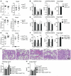
Loss of IFN-I signaling aggravates the pathology of pneumococcal pneumonia. WT and Ifnar1/ mice were infected i.n. with 105 CFU S. pneumoniae (ATCC 6303) and sacrificed at 8, 16, or 40 h p.i. (A) Bacterial load was assessed in the lungs and blood. (B) Cytokines of homogenized organs (left panel) or plasma (right panel) were quantified by ELISA. (C) Differential cell counts of BALF cells were done on cytospin preparations. (D) H&E staining of lung tissue and histology score of the staining on the right. (E) TUNEL staining of WT (left panel) and Ifnar1/ lungs (right panel) 40 h p.i. and quantification of TUNEL+ cells per field (lower panel). (F) Lung weight in WT and Ifnar1/ mice at 8, 16, and 40 h p.i. (G) Protein levels in the BALF at 8 and 16 h p.i. Data from one experiment are shown as mean + SD and are representative for two independent experiments, with eight mice per group per experiment. Two-sided t-test was performed to test for statistical significance. *p ≤ 0.05, **p ≤ 0.01, ***p ≤ 0.001, ****p ≤ 0.0001. Images in (D) and (E) are representative of two independent experiments, with eight mice per group per experiment; (10x magnification (D) and 40x (E)).
Figure 2
Figure 2
IFN-I acting on AECII prevents bacterial dissemination and damage during pneumococcal pneumonia. Ifnarfl/fl, Ifnarfl/fl Lysm-Cre+, Ifnarfl/fl Cd11c-Cre+, or Ifnarfl/fl Sftpc-CreER+ mice were infected i.n. with 105 CFU S. pneumoniae (ATCC 6303). (A–H) At 40 h p.i., the (A, C, and E) bacterial loads in lungs and blood and (B, D, and F) cytokine levels in lung, plasma, and lung weight were assessed for Ifnarfl/fl, in comparison against (A and B) Ifnarfl/fl Lysm-Cre+, (C and D) Ifnarfl/fl Cd11c-Cre+, or (E and F) Ifnarfl/fl Sftpc-CreER+ mice. (F and G) Ifnarfl/fl and Ifnarfl/fl Sftpc-CreER+ mice were injected with 5 mg Tx on four consecutive days and infected 10 days after the last Tx injection. (G) Images (upper panels) represent H&E staining of lung tissue sections at 40 h p.i. (20x magnification). Histology score (lower panel). Images are representative of two independent experiments, with six mice per group per experiment. (H) Protein concentrations were quantified in the BALF at 16 h p.i. Data from one experiment are shown as mean + SD and are representative for two independent experiments, with eight mice per group per experiment. Two-sided t-test was performed to test for statistical significance. *p ≤ 0.05, **p ≤ 0.01, ***p ≤ 0.001, ****p ≤ 0.0001.
Figure 3
Figure 3
AMs upregulate IFN-I and factors necessary for IFN-I expression upon S. pneumoniae infection. (A and B) WT mice were infected i.n. with 105 CFU S. pneumoniae (ATCC 6303) (A) IFN-β was quantified in lung homogenates by ELISA at the indicated time points. (B) BALF was taken 3 h p.i., AMs were sorted by flow cytometry, and qRT-PCR array was performed on the extracted RNA (AMs were pooled from three mice per group) (C) As in Figure 3B, AM of infected WT mice were sorted by flow cytometry and qRT-PCR for the indicated genes was performed on the extracted RNA. (D) Mice were treated i.n. with 50 μL Clodronate-filled liposomes, NaCl-filled liposomes, or NaCl 2 days prior to infection with 105 CFU S. pneumoniae (ATCC 6303). Lungs were harvested at indicated timepoints and IFN-β levels were assessed by ELISA. (E) Mice were infected with 107 CFU HI S. pneumoniae or DNAse-treated HI S. pneumoniae (both D39). Forty hours p.i. lung homogenates of WT mice were analyzed for IFN-β by ELISA (left panel), and for Ifit1 (middle panel) and Eif2ak2 (right panel) by qRT-PCR. Dashed lines (A, D, and E) indicate the detection limit. Data are shown as mean + SD. The qRT-PCR array in (B) was performed once with three mice per experiment and all depicted genes were validated by qRT-PCR in an independent experiment (C) with three mice per experiment. In (A, D, and E), data from one experiment are shown and are representative for two experiments with eight mice per group per experiment. In (D and E) one-way ANOVA followed by Tukey’s multiple comparisons analysis was performed to test for statistical significance. *p ≤ 0.05, **p ≤ 0.01, ***p ≤ 0.001, ****p ≤ 0.0001.
Figure 4
Figure 4
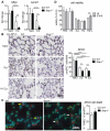
IFN-I signaling maintains AECII numbers and prevents AECII cell death upon pneumococcal pneumonia. WT and Ifnar1/ mice were infected i.n. with 105 CFU S. pneumoniae (ATCC 6303) or with 107 CFU HI S. pneumoniae (ATCC 6303; n ≥ 8 for each group). (A) Forty hours p.i. Sftpc (left panel) and Lpcat1 (right panel) levels were quantified by qRT-PCR from lung homogenates, (B) paraffin-embedded lung tissue from WT mice (left panel) and Ifnar1/ mice (right panel) was stained for proSP-C, and (C) costained for proSP-C and TUNEL; yellow arrows indicate proSP-C+ TUNEL cells and white arrows indicate pro-SP-C+ TUNEL+ cells. Quantifications of the histology are shown on the right in (B and C), respectively. (D) Serum-starved MLE cells were pretreated with 1 ng/mL IFN-β for 30 min and subsequently stimulated with 10 ng/mL pneumolysin (Ply), 5 × 10⁵ CFU/mL S. pneumoniae ATCC 6303 (S.p.), 107 CFU/mL HI S. pneumoniae ATCC 6303 (HI), or 5 μg/mL TNF-α. After 4 h cell survival was measured. Images and quantification in (B and C) are representative of two independent experiments, with eight mice per group per experiment (40x), In vitro stimulations were performed in quadruplicates. Data from one experiment are shown as mean + SD and are representative for two experiments with eight mice per group per experiment. Two-sided t-test was performed to test for statistical significance. *p ≤ 0.05, **p ≤ 0.01, ***p ≤ 0.001, ****p s 0.0001.
Figure 5
Figure 5
Absence of IFN-I signaling is linked to reduced AECII numbers upon acid induced lung injury. HCl was instilled i.t. into WT and Ifnar1/ mice and all mice were sacrificed 8 h later. (A) Lung weight (left panel) and protein concentrations (right panel) in BALF were assessed. (B) Some mice received Evans Blue i.v. 60 min before BALF and lungs were collected. Evans Blue was quantified in BALF (left panel) and formamide-pretreated lungs (right panel). (C) Sftpc (left panel) and Lpcat1 (right panel) were quantified by qRT-PCR from lung tissues. (D) Paraffin-embedded lung tissue was stained for proSP-C and (E) costained for proSP-C and TUNEL; yellow arrows indicate proSP-C+ TUNEL cells and white arrows indicate pro-SP-C+ TUNEL+ cells. Quantifications of the histology are shown on the right. Images and quantification in (C and E) are representative of two independent experiments, with eight mice per group per experiment (40x). (F) Lung homogenates were analyzed for IFN-β by ELISA and for Tnfa by qRT-PCR. Dashed lines indicate the detection limit. In vitro stimulations were performed in quadruplicates. Data from one experiment are shown as mean + SD and are representative for two experiments with eight mice per group per experiment. Two-sided t-test or one-way ANOVA followed by Tukey’s multiple comparisons analysis was performed to test for statistical significance. *p ≤ 0.05, **p ≤ 0.01, ***p ≤ 0.001, ****p ≤ 0.0001.
Figure 6
Figure 6
Proposed model of IFN-I mediated protective effects on AECII. Upon infection, AMs encounter and ingest S. pneumoniae to then induce an inflammatory response that involves secretion of IFN-I. In a similar manner, sterile ALI is associated with tissue damage and resulting inflammation. Respiratory epithelial cells are damaged in the course of pulmonary inflammation, which is diminished in the presence of IFN-I, which directly protects AECII from death.

References

    1. Trinchieri G. Type I interferon: friend or foe? J. Exp. Med. 2010;207:2053–2063. - PMC - PubMed
    1. Ivashkiv LB, Donlin LT. Regulation of type I interferon responses. Nat. Rev. Immunol. 2014;14:36–49. - PMC - PubMed
    1. Carrero JA, Calderon B, Unanue ER. Lymphocytes are detrimental during the early innate immune response against Listeria monocytogenes. J. Exp. Med. 2006;203:933–940. - PMC - PubMed
    1. Qiu H, Fan Y, Joyee AG, Wang S, Han X, Bai H, Jiao L, et al. Type I IFNs enhance susceptibility to Chlamydia muridarum lung infection by enhancing apoptosis of local macrophages. J. Immunol. 2008;181:2092–2102. - PubMed
    1. Rothfuchs AG, Trumstedt C, Mattei F, Schiavoni G, Hidmark A, Wigzell H, Rottenberg ME. STAT1 regulates IFN-alpha beta- and IFN-gamma-dependent control of infection with Chlamydia pneumoniae by nonhemopoietic cells. J. Immunol. 2006;176:6982–6990. - PubMed

Publication types

MeSH terms

Substances

LinkOut - more resources