

Neurovascular contributions to migraine: Moving beyond vasodilation
- PMID: 27312704
- PMCID: PMC5083225
- DOI: 10.1016/j.neuroscience.2016.06.012
Neurovascular contributions to migraine: Moving beyond vasodilation
Abstract
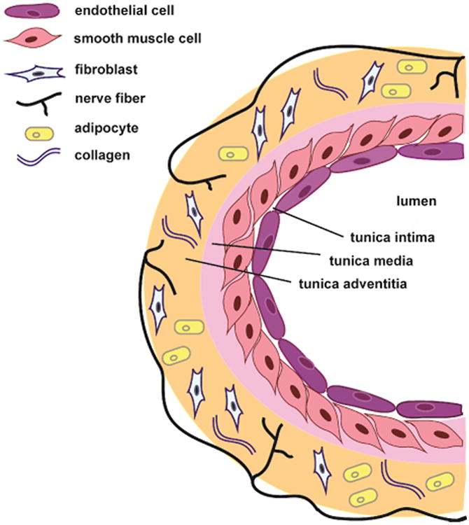
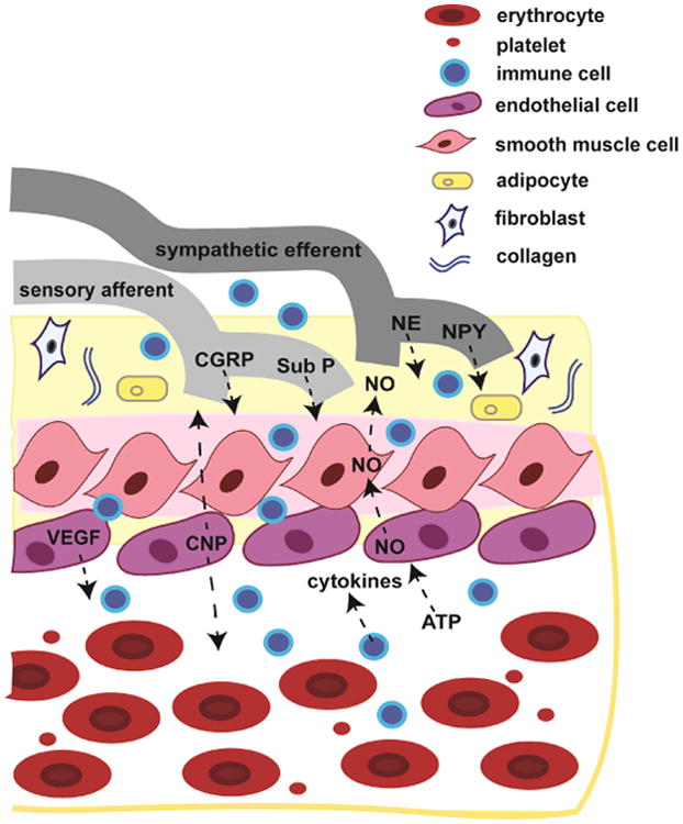
Migraine is the third most common disease worldwide, the most common neurological disorder, and one of the most common pain conditions. Despite its prevalence, the basic physiology and underlying mechanisms contributing to the development of migraine are still poorly understood and development of new therapeutic targets is long overdue. Until recently, the major contributing pathophysiological event thought to initiate migraine was cerebral and meningeal arterial vasodilation. However, the role of vasodilation in migraine is unclear and recent findings challenge its necessity. While vasodilation itself may not contribute to migraine, it remains possible that vessels play a role in migraine pathophysiology in the absence of vasodilation. Blood vessels consist of a variety of cell types that both release and respond to numerous mediators including growth factors, cytokines, adenosine triphosphate (ATP), and nitric oxide (NO). Many of these mediators have actions on neurons that can contribute to migraine. Conversely, neurons release factors such as norepinephrine and calcitonin gene-related peptide (CGRP) that act on cells native to blood vessels. Both normal and pathological events occurring within and between vascular cells could thus mediate bi-directional communication between vessels and the nervous system, without the need for changes in vascular tone. This review will discuss the potential contribution of the vasculature, specifically endothelial cells, to current neuronal mechanisms hypothesized to play a role in migraine. Hypothalamic activity, cortical spreading depression (CSD), and dural afferent input from the cranial meninges will be reviewed with a focus on how these mechanisms can influence or be impacted by blood vessels. Together, the data discussed will provide a framework by which vessels can be viewed as important potential contributors to migraine pathophysiology, even in light of the current uncertainty over the role of vasodilation in this disorder.
Keywords: cortical spreading depression; endothelial cells; hypothalamus; meningeal afferents; migraine; vasodilation.
Copyright © 2016 IBRO. Published by Elsevier Ltd. All rights reserved.
Figures
References
-
- Abdel-Samad D, Perreault C, Ahmarani L, Avedanian L, Bkaily G, Magder S, D'Orleans-Juste P, Jacques D. Differences in neuropeptide Y-induced secretion of endothelin-1 in left and right human endocardial endothelial cells. Neuropeptides. 2012;46:373–382. - PubMed
-
- Afridi SK, Giffin NJ, Kaube H, Friston KJ, Ward NS, Frackowiak RS, Goadsby PJ. A positron emission tomographic study in spontaneous migraine. Arch Neurol. 2005;62:1270–1275. - PubMed
-
- Aitken PG, Tombaugh GC, Turner DA, Somjen GG. Similar propagation of SD and hypoxic SD-like depolarization in rat hippocampus recorded optically and electrically. J Neurophysiol. 1998;80:1514–1521. - PubMed
-
- Amin FM, Asghar MS, Guo S, Hougaard A, Hansen AE, Schytz HW, van der Geest RJ, de Koning PJ, Larsson HB, Olesen J, Ashina M. Headache and prolonged dilatation of the middle meningeal artery by PACAP38 in healthy volunteers. Cephalalgia. 2012;32:140–149. - PubMed
Publication types
MeSH terms
Grants and funding
LinkOut - more resources
Full Text Sources
Other Literature Sources
Medical
Research Materials
