. 2016 Jun 17:6:28276.
doi: 10.1038/srep28276.

Single-domain flavoenzymes trigger lytic polysaccharide monooxygenases for oxidative degradation of cellulose

Affiliations

Single-domain flavoenzymes trigger lytic polysaccharide monooxygenases for oxidative degradation of cellulose

Sona Garajova et al. Sci Rep. .

Abstract

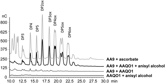
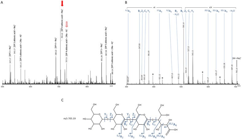
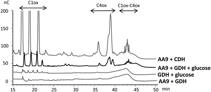
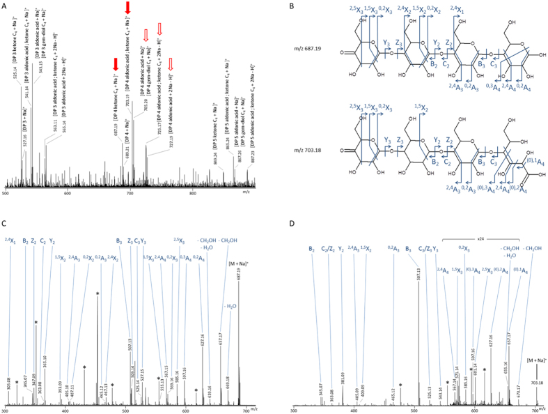
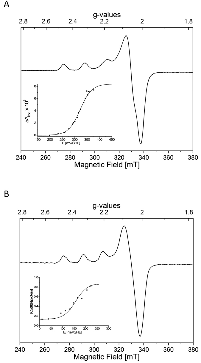
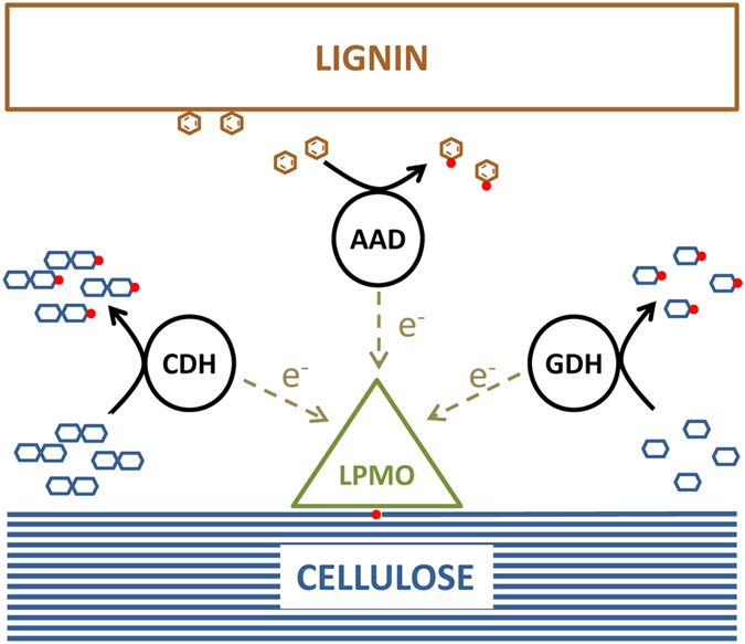
The enzymatic conversion of plant biomass has been recently revolutionized by the discovery of lytic polysaccharide monooxygenases (LPMOs) that carry out oxidative cleavage of polysaccharides. These very powerful enzymes are abundant in fungal saprotrophs. LPMOs require activation by electrons that can be provided by cellobiose dehydrogenases (CDHs), but as some fungi lack CDH-encoding genes, other recycling enzymes must exist. We investigated the ability of AA3_2 flavoenzymes secreted under lignocellulolytic conditions to trigger oxidative cellulose degradation by AA9 LPMOs. Among the flavoenzymes tested, we show that glucose dehydrogenase and aryl-alcohol quinone oxidoreductases are catalytically efficient electron donors for LPMOs. These single-domain flavoenzymes display redox potentials compatible with electron transfer between partners. Our findings extend the array of enzymes which regulate the oxidative degradation of cellulose by lignocellulolytic fungi.


Figures

Figure 1
Figure 1. Analysis of degradation products generated by PaLPMO9E in the presence of aryl-alcohol dehydrogenase (AAQO1).
HPAEC chromatograms of the oligosaccharides released upon degradation of 0.1% PASC with 4.4 μM LPMO in the presence of 4.4 μM AAQO1 and 2.5 mM of anisyl alcohol, at 30 °C for 24 h. The positive control was obtained by degradation of 0.1% PASC with 4.4 μM PaLPMO9E in the presence of 1 mM ascorbate, at 30 °C for 24 h.
Figure 2
Figure 2. Mass spectrometry analysis of degradation products generated from PASC by AA9 LPMO with AAQO1 and anisyl alcohol as described in Fig. 1.
Analyses were performed after 24 hours of cellulose degradation. Panel (A) shows MS spectrum of sample with peaks corresponding to native and oxidized cello-oligosaccharides. Peaks that were further fragmented are indicated by arrows. Panel (B) shows the MS/MS spectrum of the 705 m/z species which corresponds to a C1-oxidized product. Observed fragments are depicted on the structure in panel (C). Black stars: unassigned fragments.
Figure 3
Figure 3. Analysis of degradation products generated from cellulose by PaLPMO9H in the presence of GDH.
Chromatograms of the oligosaccharides released upon degradation of 0.1% PASC with 4.4 μM LPMO in the presence of 4.4 μM GDH and 0.5 mM glucose at 40 °C for 24 h. The positive control was obtained by degradation of 0.1% PASC with 4.4 μM LPMO and 1.4 μM of CDH, at 40 °C for 24 h.
Figure 4
Figure 4. Mass spectrometry analysis of degradation products generated from PASC by PaLPMO9H with GDH and glucose as described in Fig. 3.
Analyses were performed after 24 hours of cellulose degradation. Panel (A) shows MS spectrum of sample with peaks corresponding to native and oxidized cello-oligosaccharides. Peaks that were further fragmented are indicated by arrows. Panel (B) shows MS/MS spectrum of the 687 m/z species which corresponds to a C4-oxidized product (ketone form). Panel (C) shows MS/MS spectrum of the 703 m/z species which corresponds to a double oxidized product (ketone form on C4 and aldonic acid form on C1). Observed fragments are depicted on structures in panel (D). Black stars: unassigned fragments.
Figure 5
Figure 5
X-band EPR spectra of (A) PaLPMO9H 300 μM and (B) PaLPMO9E 145 μM at pH 5. Spectra were recorded at 50 K, 1 mW microwave power at 9.480 GHz, and 3 mT modulation amplitude. Titration curves are given in insets.
Figure 6
Figure 6. Proposed scheme of fungal synergies for oxidative degradation of cellulose.
Products of cellulose and lignin degradation are substrates for fungal dehydrogenases (CDH, GDH and AAQO) which provide electrons to LPMOs.

References

    1. Harris P. V. et al. Stimulation of lignocellulosic biomass hydrolysis by proteins of glycoside hydrolase family 61: Structure and function of a large, enigmatic family. Biochemistry 49, 3305–3316 (2010). - PubMed
    1. Vaaje-Kolstad G. et al. An oxidative enzyme boosting the enzymatic conversion of recalcitrant polysaccharide. Science 330, 219–222 (2010). - PubMed
    1. Johansen K. S. Discovery and industrial applications of lytic polysaccharide mono-oxygenases. Biochem. Soc. Trans. 44, 143–149 (2016). - PubMed
    1. Lombard V., Golaconda Ramulu H., Drula E., Coutinho P. M. & Henrissat B. The carbohydrate-active enzymes database (CAZy) in 2013. Nucleic Acids Res. 42, 490–495 (2014). - PMC - PubMed
    1. Quinlan R. J. et al. Insights into the oxidative degradation of cellulose by a copper metalloenzyme that exploits biomass components. Proc. Natl. Acad. Sci. USA 108, 15079–84 (2011). - PMC - PubMed

Publication types

MeSH terms

LinkOut - more resources