. 2016 Jun 17;352(6292):1474-7.
doi: 10.1126/science.aad5168.

Biosensor reveals multiple sources for mitochondrial NAD⁺

Affiliations

Biosensor reveals multiple sources for mitochondrial NAD⁺

Xiaolu A Cambronne et al. Science. .

Abstract

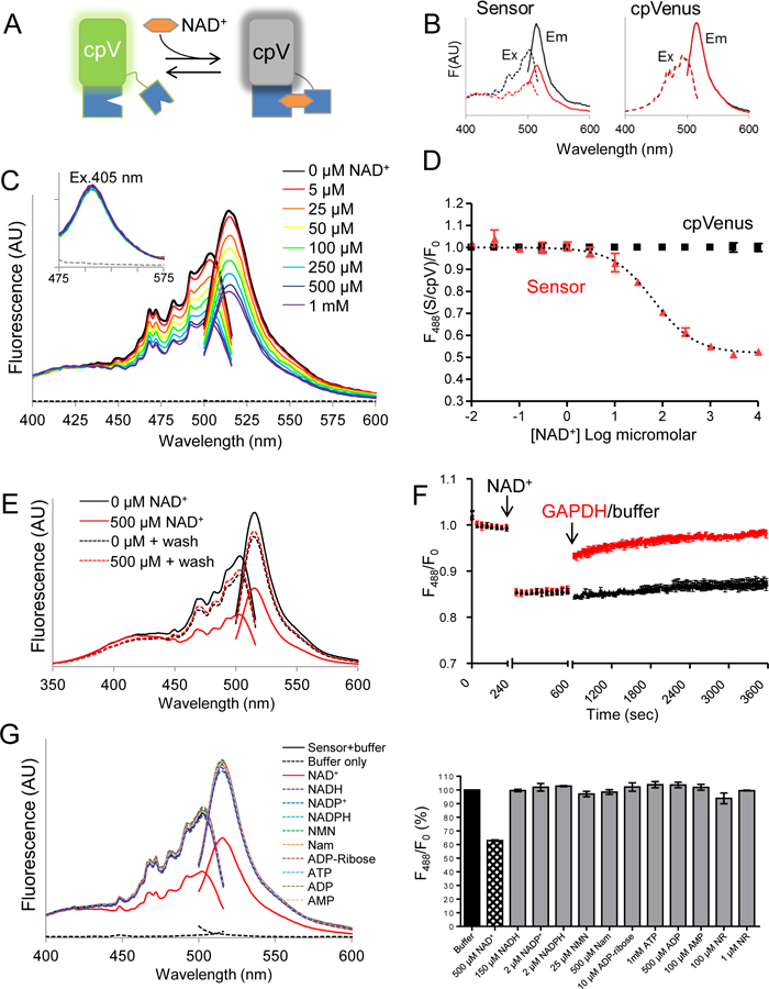
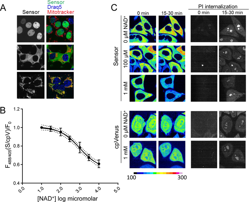
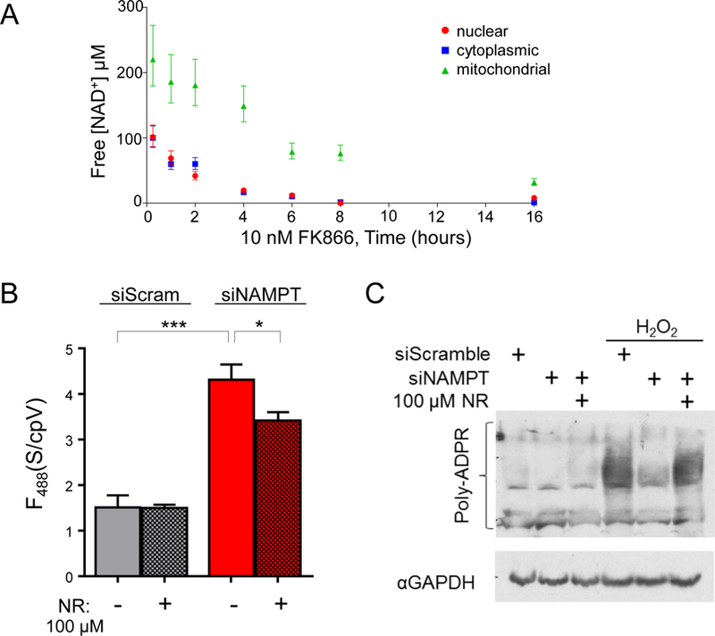
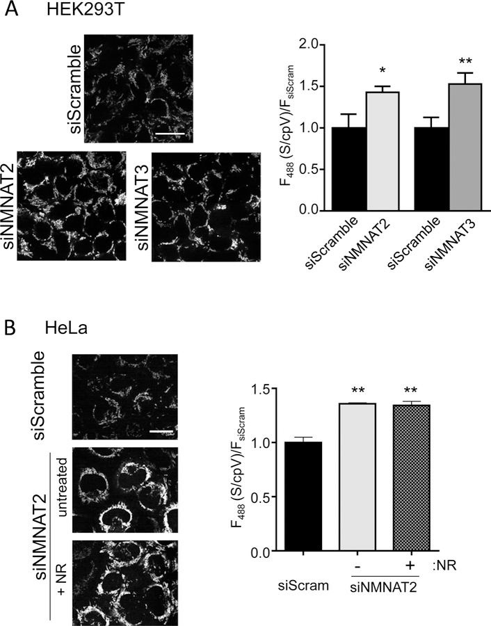
Nicotinamide adenine dinucleotide (NAD(+)) is an essential substrate for sirtuins and poly(adenosine diphosphate-ribose) polymerases (PARPs), which are NAD(+)-consuming enzymes localized in the nucleus, cytosol, and mitochondria. Fluctuations in NAD(+) concentrations within these subcellular compartments are thought to regulate the activity of NAD(+)-consuming enzymes; however, the challenge in measuring compartmentalized NAD(+) in cells has precluded direct evidence for this type of regulation. We describe the development of a genetically encoded fluorescent biosensor for directly monitoring free NAD(+) concentrations in subcellular compartments. We found that the concentrations of free NAD(+) in the nucleus, cytoplasm, and mitochondria approximate the Michaelis constants for sirtuins and PARPs in their respective compartments. Systematic depletion of enzymes that catalyze the final step of NAD(+) biosynthesis revealed cell-specific mechanisms for maintaining mitochondrial NAD(+) concentrations.


Figures

Fig. 1
Fig. 1. Characterization of an NAD+ biosensor.
(A) The NAD+ biosensor comprises cpVenus (cpV) and a bipartite NAD+-binding domain (blue). The unbound species fluoresces following excitation at 488 nm; NAD+ binding causes a loss of fluorescence. (B) Excitation (dashed lines) and emission (solid lines) scans of purified sensor with either 0 µM (black) or 500 µM (red) NAD+. Excitation was monitored at 530 nm and emission was monitored after excitation at 488 nm. (C) Fluorescence emission and excitation scans at indicated NAD+ concentrations or buffer only control (dashed lines). Inset shows fluorescence from excitation at 405 nm. (D) Maxima from 488 nm emission peaks of sensor and cpVenus (250 nM) at indicated NAD+ concentrations; mean±SD, n=3. (E) Fluorescence excitation and emission of sensor incubated with 0 µM (black solid) or 500 µM NAD+ (red solid). NAD+ was washed out and fluorescence was re-evaluated in each sample (dotted lines). (F) GAPDH (red) increases sensor fluorescence monitored at 520 nm following excitation at 488 nm. (G) (left) Excitation and emission profiles and (right) maxima from 488 nm emission with indicated substrates. mean±SD, n=3.
Fig. 2
Fig. 2. Free intracellular NAD+ measurements.
(A) HEK293T cells stably expressing nuclear, cytoplasmic, or mitochondrial sensors. Nuclear marker Draq5 (blue), mitochondrial marker Mitotracker CMXRos (red), sensor (green). (B) Cytoplasmic sensor (S) was calibrated for NAD+-dependent fluorescence changes in digitonin-permeabilized HEK293T cells. Ratio of 488/405 nm fluorescence measured with flow cytometry were normalized to cpVenus (cpV) and fit with a variable slope model; 95% confidence interval (dotted lines). (C) Representative images from adherent HeLa cells permeabilized with saponin in the presence of indicated NAD+ concentrations, as monitored by propidium iodide (PI) internalization, ex. 561, em. 617/73 (right). Live images were captured every 2.5 minutes and fluorescence intensity from 488 nm excitation is normalized to the indicated scale bar.
Fig. 3
Fig. 3. NAD+ fluctuations in cells.
(A) Free NAD+ concentrations in HEK293T cells following treatment with FK866 (10 nM). Fluorescence of Sensor/cpVenus (488/405 nm) were measured by flow cytometry and the fold- change compared to untreated controls was interpolated onto an in vitro standard curve. (B) Cytoplasmic free NAD+ was decreased by NAMPT depletion and partially restored by NR treatment (100 µM). Imaging measurements from ex. 488 nm. REML, mean±SEM, n=3, ***p<0.001, *p<0.05. (C) Effect of free NAD+ availability on PARP activation.
Fig. 4
Fig. 4. Multiple sources for mitochondrial NAD+.
(A) Imaging measurements of mitochondrial NAD+ sensor after depletion of either NMNAT2 or NMNAT3 in HEK293T cells. Scale bar, 25 μm. Ex. 488 nm fluorescence was normalized to cpVenusMito. Mean±SEM, n=3, REML, *p=0.03, **p<0.005. (B) Effect of NR treatment (250 µM, 24 hours) on NAD+ levels in the mitochondria of HeLa cells depleted of NMNAT2. Representative images, left. Scale bar, 25 μm. Quantitation of fluorescence, right, relative to cpVenusMito and siScramble. Mean±SEM, n=3, REML, **p<0.01.

Comment in

References

    1. Cantó C, Menzies KJ, Auwerx J. NAD(+) Metabolism and the Control of Energy Homeostasis: A Balancing Act between Mitochondria and the Nucleus. Cell Metab 22, 31–53 (2015). - PMC - PubMed
    1. Sauve AA, Celic I, Avalos J, Deng H, Boeke JD, et al. Chemistry of gene silencing: the mechanism of NAD+-dependent deacetylation reactions. Biochemistry 40, 15456–15463 (2001). - PubMed
    1. Hassa PO, Haenni SS, Elser M, Hottiger MO. Nuclear ADP-ribosylation reactions in mammalian cells: where are we today and where are we going? Microbiol Mol Biol Rev 70, 789–829 (2006). - PMC - PubMed
    1. Revollo JR, Grimm AA, Imai S. The NAD biosynthesis pathway mediated by nicotinamide phosphoribosyltransferase regulates Sir2 activity in mammalian cells. J Biol Chem 279, 50754–50763 (2004). - PubMed
    1. Berger F, Lau C, Dahlmann M, Ziegler M. Subcellular compartmentation and differential catalytic properties of the three human nicotinamide mononucleotideadenylyltransferase isoforms. J Biol Chem 280, 36334–36341 (2005). - PubMed

Publication types

MeSH terms