. 2016 Jul 5;113(27):E3931-40.
doi: 10.1073/pnas.1600428113. Epub 2016 Jun 16.

VgrG C terminus confers the type VI effector transport specificity and is required for binding with PAAR and adaptor-effector complex

Affiliations

VgrG C terminus confers the type VI effector transport specificity and is required for binding with PAAR and adaptor-effector complex

Devanand D Bondage et al. Proc Natl Acad Sci U S A. .

Abstract

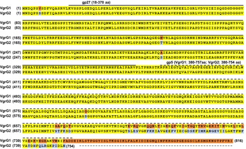
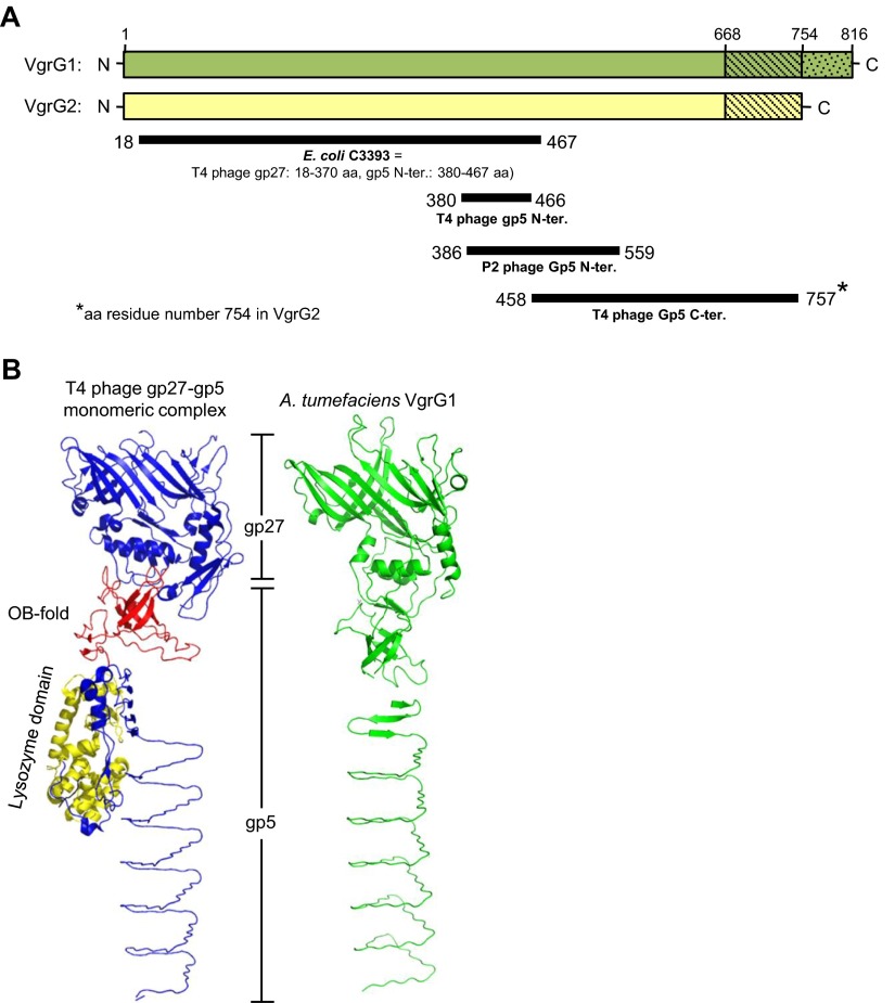
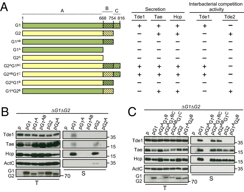
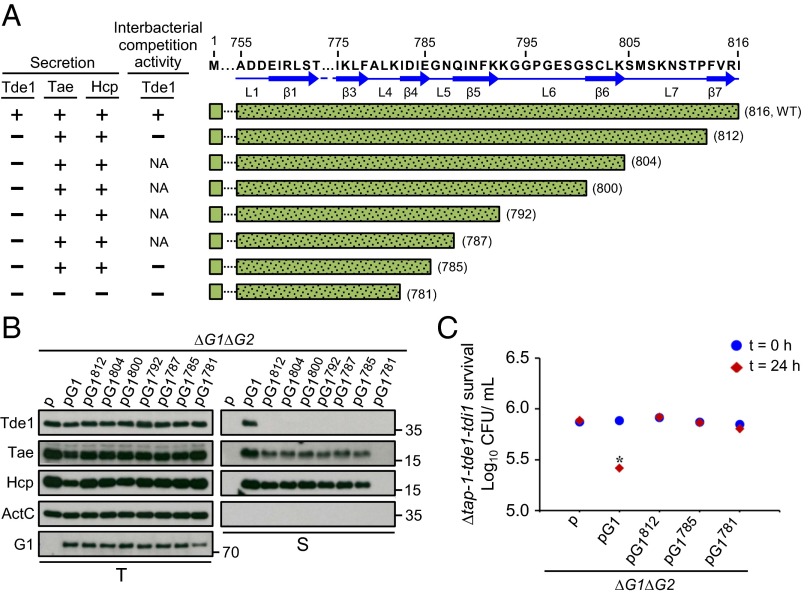
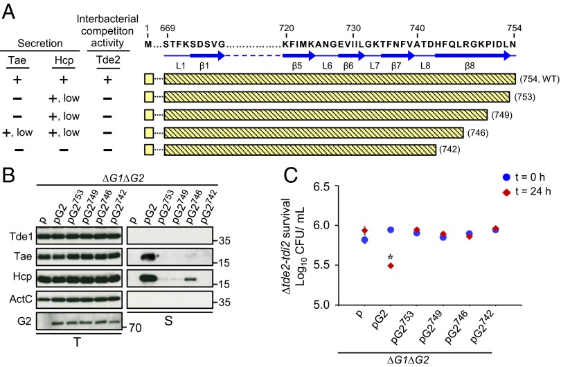
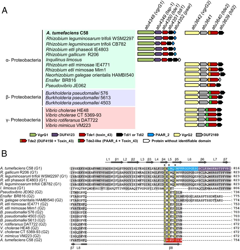
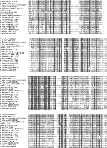
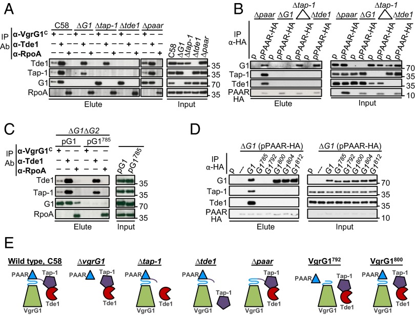
Type VI secretion system (T6SS) is a macromolecular machine used by many Gram-negative bacteria to inject effectors/toxins into eukaryotic hosts or prokaryotic competitors for survival and fitness. To date, our knowledge of the molecular determinants and mechanisms underlying the transport of these effectors remains limited. Here, we report that two T6SS encoded valine-glycine repeat protein G (VgrG) paralogs in Agrobacterium tumefaciens C58 specifically control the secretion and interbacterial competition activity of the type VI DNase toxins Tde1 and Tde2. Deletion and domain-swapping analysis identified that the C-terminal extension of VgrG1 specifically confers Tde1 secretion and Tde1-dependent interbacterial competition activity in planta, and the C-terminal variable region of VgrG2 governs this specificity for Tde2. Functional studies of VgrG1 and VgrG2 variants with stepwise deletion of the C terminus revealed that the C-terminal 31 aa (C31) of VgrG1 and 8 aa (C8) of VgrG2 are the molecular determinants specifically required for delivery of each cognate Tde toxin. Further in-depth studies on Tde toxin delivery mechanisms revealed that VgrG1 interacts with the adaptor/chaperone-effector complex (Tap-1-Tde1) in the absence of proline-alanine-alanine-arginine (PAAR) and the VgrG1-PAAR complex forms independent of Tap-1 and Tde1. Importantly, we identified the regions involved in these interactions. Although the entire C31 segment is required for binding with the Tap-1-Tde1 complex, only the first 15 aa of this region are necessary for PAAR binding. These results suggest that the VgrG1 C terminus interacts sequentially or simultaneously with the Tap-1-Tde1 complex and PAAR to govern Tde1 translocation across bacterial membranes and delivery into target cells for antibacterial activity.

Keywords: Agrobacterium tumefaciens; DNase effector; VgrG; interbacterial competition; type VI secretion system.


Conflict of interest statement

The authors declare no conflict of interest.

Figures

Fig. S1.
Fig. S1.
Amino acid sequence alignment of A. tumefaciens C58 VgrG1 and VgrG2. Identical residues are in yellow and variable residues in VgrG1 and VgrG2 are in orange and light blue, respectively. The solid and dashed lines represent regions with predicted gp27 and gp5 domains, respectively. Number in brackets at both sides of sequences is the residue number.
Fig. S2.
Fig. S2.
Domain prediction and structure modeling of VgrG1 and VgrG2. (A) Graphic representation of the domains predicted to present in VgrG1 and VgrG2 by the Phyre2 web server. VgrG1 and VgrG2 full-length proteins are represented as green and yellow bars, respectively (drawn to scale). Solid dark lines represent the region of VgrG1/VgrG2 that is structurally similar (confidence > 99%) to the protein shown in bold under each line. The number at the start and end of each line indicates the amino acid position from VgrG1/VgrG2. According to ref. , E. coli C3393 protein is structurally similar to the gp27 protein and the N-terminal of gp5 protein in T4 phage. (B) Structure comparison of A. tumefaciens VgrG1 and phage gp27-gp5 monomeric complex derived from the gp5–gp27 trimeric complex (PDB ID code 1K28). Shown for VgrG1 the best-predicted model among nine different models generated using PHYRE2, SWISS-MODEL, and I-TAESSER homology modeling servers. In the gp27–gp5 monomeric complex structure, the gp27 domain and gp5 C terminus is shown in blue, the OB fold in red and the lysozyme domain in yellow. The predicted structure of A. tumefaciens VgrG1 is in green.
Fig. 1.
Fig. 1.
Genetic organization of vgrG1 and vgrG2 and their requirement in specific Tde toxin delivery. (A) Genetic organization of vgrG2 and hcp operons of A. tumefaciens strain C58. The genes are indicated by the annotated locus tag or designated name adapted from the literature (6, 37). The vgrG genes are in blue and the genes encoding the three toxins and their cognate immunity proteins are in red and green, respectively. (B) VgrG1 is required for secretion of Tde1 but not Tae. Western blot analysis of the total (T) and secreted (S) proteins from various A. tumefaciens strains expressing the plasmid control (p) or indicated plasmid. Protein names and molecular weight markers are indicated at the left and right, respectively. The soluble ActC protein was used as an internal nonsecreted protein control. (C and D) VgrG1 and VgrG2 are required for Tde1- and Tde2-dependent interbacterial competition activity, respectively. Various A. tumefaciens strains shown on the x axis were each mixed with ∆atu4349-tde1-tdi1 (C) or ∆tde2-tdi2 (D) at a 10:1 ratio and infiltrated into N. benthamiana leaves. The survival of ∆atu4349-tde1-tdi1 and ∆tde2-tdi2 collected at 0 and 24-h postinoculation (h) was quantified as cfu. Data are mean ± SEM (C, n = 4 biological repeats from two independent experiments; D, n = 5 biological repeats from three independent experiments). Significant difference compared with WT C58 at 24-h postinfiltration (*P ≤ 0.01). vgrG1 and vgrG2 are abbreviated as G1 and G2, respectively.
Fig. 2.
Fig. 2.
C-terminal variable/extension region of VgrG is responsible for Tde toxin secretion and interbacterial competition activity. (A) Schematic representation of full-length and truncated VgrG1 (green) and VgrG2 (yellow). Highly conserved region (1–668 aa), variable region (669–754 aa), and VgrG1 C-terminal extension (755–816 aa) are denoted as regions A, B, and C, respectively. The name of each variant is indicated at the left. The presence (+) or absence (−) of Tde1, Hcp, and Tae secretion is based on B and C. Presence (+) and absence (–) of Tde1- and Tde2-dependent antibacterial activity is based on Fig. S3. (B) Effect of VgrG variable and C-terminal extension deletion and (C) chimeric VgrG variants on type VI secretion. Western blot analysis of total (T) and secreted (S) protein from A. tumefaciensvgrG1vgrG2 expressing the plasmid control (p) or indicated plasmid. Protein names and molecular weight markers are indicated at the left and right, respectively. The soluble ActC protein was used as an internal nonsecreted protein control. vgrG1 and vgrG2 are abbreviated as G1 and G2, respectively.
Fig. S3.
Fig. S3.
Effect of truncated and chimeric VgrG variants on (A and C) Tde1- and (B and D) Tde2-dependent interbacterial competition activity. Various A. tumefaciens strains shown on the x axis were mixed with ∆tap-1-tde1-tdi1 (A and C) or ∆tde2-tdi2 (B and D) at a 10:1 ratio and infiltrated into N. benthamiana leaves. The survival of ∆tap-1-tde1-tdi1 and tde2-tdi2 at 0 and 24-h postinfiltration was quantified as cfu. Data are mean ± SEM (n = 3 biological repeats for A and n = 4 biological repeats for B–D from two independent experiments). Significant difference compared with ∆vgrG1vgrG2 (∆G1G2) at 24-h postinfiltration (*P ≤ 0.01). vgrG1 and vgrG2 are abbreviated as G1 and G2, respectively.
Fig. 3.
Fig. 3.
Effect of VgrG1 C-terminal truncation on type VI secretion and Tde1-dependent interbacterial competition activity. (A) Amino acid sequence of VgrG1 C-terminal extension shown with the indicated residue number and predicted β-strands and loops marked with blue solid arrows and lines, respectively. VgrG1 C-terminal truncated variants are shown with green bars filled with dots, and the number in parentheses represents the terminal amino acid of each variant. Presence (+) or absence (−) of Tde1, Hcp, and Tae secretion and Tde1-dependent antibacterial activity is based on Fig. 4 B and C. NA: not analyzed. (B) Western blot analysis of total (T) and secreted (S) proteins from A. tumefaciensvgrG1vgrG2 expressing the plasmid control (p) or indicated plasmid. Protein names and molecular weight markers are at the left and right, respectively. The soluble ActC protein was used as an internal nonsecreted protein control. (C) Effect of VgrG1 C-terminal truncation on Tde1-dependent interbacterial competition activity. Various A. tumefaciens strains shown on the x axis were mixed with ∆tap-1-tde1-tdi1 at a 10:1 ratio and infiltrated into N. benthamiana leaves. The survival of ∆tap-1-tde1-tdi1 collected at 0 and 24 h was quantified as cfu. Data are mean ± SEM (n = 5 biological repeats from two independent experiments). Significant difference compared with ∆vgrG1vgrG2 (∆G1G2) at 24-h postinfiltration (*P ≤ 0.01). vgrG1 and vgrG2 are abbreviated as G1 and G2, respectively.
Fig. 4.
Fig. 4.
Effect of VgrG2 C-terminal truncation on type VI secretion and Tde2-dependent interbacterial competition activity. (A) Amino acid sequence of VgrG2 C-terminal variable region is shown with the indicated residue number, and the predicted β-strands and loops are marked with blue solid arrows and lines, respectively. VgrG2 C-terminal truncated variants are shown with yellow bars, and the numbers in parentheses represent the terminal amino acid of each variant. Presence (+) or absence (–) of Hcp and Tae secretion and Tde2-dependent antibacterial activity is based on Fig. 3 B and C. (B) Western blot analysis of total (T) and secreted (S) protein from A. tumefaciensvgrG1vgrG2 expressing the plasmid control (p) or indicated plasmid. Protein names and molecular weight markers are indicated at the left and right, respectively. The soluble ActC protein was an internal nonsecreted protein control. (C) Effect of VgrG2 C-terminal truncation on Tde2-dependent interbacterial competition activity. Various A. tumefaciens strains shown on the x axis were mixed with ∆tde2-tdi2 at a 10:1 ratio and infiltrated into N. benthamiana leaves. The survival of ∆tde2-tdi2 collected at 0 and 24 h was quantified as cfu. Data are mean ± SEM (n = 5 biological repeats from two independent experiments). Significant difference compared with ∆vgrG1vgrG2 (∆G1G2) at 24-h postinfiltration (*P ≤ 0.01). vgrG1 and vgrG2 are abbreviated as G1 and G2, respectively.
Fig. 5.
Fig. 5.
(A) Conservation of the vgrG1-tap-1-tde1-tdi1-paar and vgrG2-atu3641-tde2-tdi2 gene clusters in Proteobacteria. Bacteria belonging to different proteobacterial classes are shaded with different colors. Genetic organization at right of each bacterium was deduced from manual inspection of respective bacterial genome and color-coded by the presence of a conserved protein domain as predicted by an NCBI-CD search algorithm (39). The locus tags/names of A. tumefaciens C58 genes are indicated and the color-coded protein domains are listed and drawn to scale. (B) C-terminal amino acid sequence alignment of tde-associated vgrG homologs. C-terminal regions of identified VgrG1 and VgrG2 homologs aligned in Fig. S5 are presented. The amino acid residues in dark gray represent highly conserved residues and in light gray, partially conserved residues. Numbers at the right represent the terminal amino acid residue of each aligned VgrG homolog. The VgrG1 C31 sequence is shaded in blue (region required for PAAR binding) and purple (region dispensable for PAAR but required for interacting with Tap-1–Tde1 complex) and the VgrG2 C8 sequence is shaded in red. A common I/LxG/AxxI/V motif located at the boundary uncoupling Hcp and Tde toxin delivery is highlighted and indicated by asterisks (*). The predicted secondary structure of A. tumefaciens C58 VgrG1 and VgrG2 is indicated at the top and bottom, respectively.
Fig. S4.
Fig. S4.
Flowchart for identification of (A) Tde1 orthologs, Tde2 orthologs, and Tde2-like proteins and (B) VgrG1 and VgrG2 orthologs.
Fig. S5.
Fig. S5.
Amino acid sequence alignment of tde-linked vgrG homologs. Identified VgrG1 and VgrG2 homologs are aligned and the amino acid residues in dark gray representing highly conserved residues or in light gray indicate partially conserved residues. Numbers at the right represent the amino acid residue positions of each aligned VgrG homolog. Gene ID of VgrG proteins from top to bottom: 15158894, 745837943, 393183390, 749199676, 574594253, 652473941, 211959592, 517049910, 659658772, 647796246, 528839901, 217394189, 714469101, 705577537, 498139720, 447019410, 487841774, 262025037, 159185839.
Fig. S5.
Fig. S5.
Amino acid sequence alignment of tde-linked vgrG homologs. Identified VgrG1 and VgrG2 homologs are aligned and the amino acid residues in dark gray representing highly conserved residues or in light gray indicate partially conserved residues. Numbers at the right represent the amino acid residue positions of each aligned VgrG homolog. Gene ID of VgrG proteins from top to bottom: 15158894, 745837943, 393183390, 749199676, 574594253, 652473941, 211959592, 517049910, 659658772, 647796246, 528839901, 217394189, 714469101, 705577537, 498139720, 447019410, 487841774, 262025037, 159185839.
Fig. 6.
Fig. 6.
The effect of tde-associated genes tap-1, paar, and atu3641 for cognate Tde effector translocation. Requirement of tap-1 (A and D) and paar genes (B and E) in Tde1-dependent antibacterial activity and Tde1 secretion; requirement of atu3641 in Tde2-dependent antibacterial activity (C) and type VI secretion (F). (A–C) E. coli killing assay in which attacker A. tumefaciens strains containing the plasmid only (p) or expressing the indicated genes shown on the x axis were mixed with E. coli DH10B cells expressing tdi1 or tdi2 at a 30:1 ratio for competition. The survival of E. coli cells was quantified as cfu and shown on the y axis. Data are mean ± SEM (n = 5 biological repeats from two independent experiments) computed by one-way ANOVA. Different letters above the bar indicate significant difference (P < 0.05) determined by Tukey’s HSD test. (D–F) Western blot analysis of the total (T) and secreted (S) proteins from various A. tumefaciens strains expressing the plasmid control (p) or indicated gene. Protein names and molecular weight markers are indicated at the left and right, respectively. The soluble ActC protein was used as an internal nonsecreted protein control.
Fig. 7.
Fig. 7.
Interactions of VgrG1, Tap-1, Tde1, and PAAR proteins. Co-IP of WT A. tumefaciens C58 and indicated mutant strains (A) or ∆vgrG1vgrG2 (∆G1G2) expressing full-length vgrG1 (G1) and with the vgrG1 C31 deletion variant (G1785) (C) by using α-VgrG1 C-terminal epitope and α-Tde1 antibodies. α-RpoA antibody was used as a negative control. Co-IP of various mutants by using strains expressing pRL662 plasmid (p) only or PAAR-HA (B) or ∆G1(pPAAR-HA) expressing full-length VgrG1 (G1) or each of truncated VgrG1 variants (D) by anti-HA antibody. The resulting total protein extract was used as input for Co-IP. Coprecipitated proteins were detected by Western blot analysis with antiserum specific to indicated proteins. Proteins in input and elute fractions were detected by Western blot analysis. Protein names and molecular weight markers are indicated at the left and right, respectively. (E) A summary model of protein–protein interactions. Each protein is color-coded and C31 of VgrG1 is presented as β-helix consisting L5-β7 (L5-β5-L6 in blue and β6-L7-β7 in purple) at tip of VgrG spike.
Fig. S6.
Fig. S6.
(A) Distribution of conserved DUF2169 domain from Atu3641 across different bacterial phyla according to Pfam database. (B) Selected bacterial species in which the DUF2169-containing protein is linked with VgrG2 orthologs and effectors other than Tde2/Tde2L. Gene ID of VgrG2 orthologs from top to bottom: 739337228, 654370902, 590115676, 741017205, 498355758, 657887367. (C) Stability of Tde2HD-Strep in the presence and absence of Atu3641-HA. Tde2HD-Strep was expressed alone or with Atu3641-HA in the A. tumefacienstde2–atu3641 deletion strain. Western blot analysis of total protein from cells grown in 523-medium. Band intensity of Tde2HD-Strep was quantified by using ImageJ software (49) and expressed as a percentage of the plasmid-only control, which was set to 0%. Data are mean ± SEM obtained from four independent experiments, ***P < 0.01. Representative blot at the bottom shows a Tde HD -Strep and Atu3641-HA protein band. 1: ∆tde2-atu3641 (p); 2: ∆tde2-atu3641 (p-Tde2HD-Strep); 3: ∆tde2-atu3641 (p-Tde2HD-Strep + Atu3641-HA).

References

    1. Durand E, Cambillau C, Cascales E, Journet L. VgrG, Tae, Tle, and beyond: The versatile arsenal of Type VI secretion effectors. Trends Microbiol. 2014;22(9):498–507. - PubMed
    1. Russell AB, et al. Type VI secretion delivers bacteriolytic effectors to target cells. Nature. 2011;475(7356):343–347. - PMC - PubMed
    1. English G, et al. New secreted toxins and immunity proteins encoded within the Type VI secretion system gene cluster of Serratia marcescens. Mol Microbiol. 2012;86(4):921–936. - PMC - PubMed
    1. Whitney JC, et al. Identification, structure, and function of a novel type VI secretion peptidoglycan glycoside hydrolase effector-immunity pair. J Biol Chem. 2013;288(37):26616–26624. - PMC - PubMed
    1. Altindis E, Dong T, Catalano C, Mekalanos J. Secretome analysis of Vibrio cholerae type VI secretion system reveals a new effector-immunity pair. MBio. 2015;6(2):e00075. - PMC - PubMed

Publication types

MeSH terms

Substances