. 2016 Jun 15;30(12):1454-69.
doi: 10.1101/gad.282400.116. Epub 2016 Jun 16.

Mechanotransduction activates canonical Wnt/β-catenin signaling to promote lymphatic vascular patterning and the development of lymphatic and lymphovenous valves

Affiliations

Mechanotransduction activates canonical Wnt/β-catenin signaling to promote lymphatic vascular patterning and the development of lymphatic and lymphovenous valves

Boksik Cha et al. Genes Dev. .

Abstract

Lymphatic vasculature regulates fluid homeostasis by returning interstitial fluid to blood circulation. Lymphatic endothelial cells (LECs) are the building blocks of the entire lymphatic vasculature. LECs originate as a homogeneous population of cells predominantly from the embryonic veins and undergo stepwise morphogenesis to become the lymphatic capillaries, collecting vessels or valves. The molecular mechanisms underlying the morphogenesis of the lymphatic vasculature remain to be fully understood. Here we show that canonical Wnt/β-catenin signaling is necessary for lymphatic vascular morphogenesis. Lymphatic vascular-specific ablation of β-catenin in mice prevents the formation of lymphatic and lymphovenous valves. Additionally, lymphatic vessel patterning is defective in these mice, with abnormal recruitment of mural cells. We found that oscillatory shear stress (OSS), which promotes lymphatic vessel maturation, triggers Wnt/β-catenin signaling in LECs. In turn, Wnt/β-catenin signaling controls the expression of several molecules, including the lymphedema-associated transcription factor FOXC2. Importantly, FOXC2 completely rescues the lymphatic vessel patterning defects in mice lacking β-catenin. Thus, our work reveals that mechanical stimulation is a critical regulator of lymphatic vascular development via activation of Wnt/β-catenin signaling and, in turn, FOXC2.

Keywords: FOXC2; PROX1; Wnt/β-catenin signaling; lymphatic valves; lymphatic vascular development; lymphovenous valves.


Figures

Figure 1.
Figure 1.
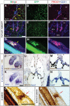
The canonical Wnt/β-catenin signaling pathway is active in the developing LVVs and LVs. (AI) TCF/LEF-H2BEGFP embryos were collected at E12.0 (AC), E14.5 (DF), and E18.5 (GI). E12.0 and E14.5 embryos were frontally sectioned, and IHC was performed for GFP, PROX1, and the pan-EC marker CD31. The mesenteries of E18.5 embryos were analyzed by whole-mount IHC for the same markers. The canonical Wnt/β-catenin signaling pathway is active in the GFP+ cells of the LVVs (AF, yellow arrows) and LVs (GI, yellow arrow). (JN) The expression of Axin2, a target of Wnt/β-catenin signaling pathway, was analyzed in E14.5 embryos by in situ hybridization. Adjacent sections were coimmunostained for PROX1. (J,K) PROX1+ ECs and the mesenchyme of the semilunar valves of the heart are Axin2+. (LN) LVVs (arrows) strongly express Axin2. (O,P) The mesenteric lymphatic vessels were immunostained for total β-catenin (O) or nonphosphorylated active β-catenin (P). LVs are strongly labeled by both antibodies (red arrows). (LS) Lymph sacs; (IJV) internal jugular vein; (SCV) subclavian vein; (SVC) superior vena cava. Bars: A–F,J–P, 100 µm; GI, 50 µm. n = 4 for each experiment.
Figure 2.
Figure 2.
β-Catenin is necessary for the development of LVVs, LVs, VVs, and cardiac valves. (AJ) The gene encoding β-catenin (Ctnnb1) was deleted from LECs, LVV-ECs, LV-ECs, and VV-ECs using Lyve1-Cre mice. IHC was performed for the indicated markers. Frontal sections from E14.5 (AD) and E16.5 (G,H) embryos revealed the LVVs in controls (arrows) but not in their Lyve1-Cre; Ctnnb1LOF littermates. Arrowheads point to the valve-forming area of mutants. (G,H) At E16.5, VVs are seen in control embryos (yellow arrows) but not in mutants. (E,F) SEM confirmed the presence of LVV-ECs (magenta) and VV-ECs (green) in E14.5 control embryos and their absence in Lyve1-Cre;Ctnnb1LOF littermates. (I,J) Whole-mount IHC of the mesenteric lymphatic vessels revealed the presence of LVs in E18.5 control (arrows) but not Lyve1-Cre;Ctnnb1LOF littermates. (I) Furthermore, PROX1 is strongly expressed in the LVs but down-regulated elsewhere in the lymphatic vessels of controls. (J) In contrast, PROX1 expression is uniformly high in the lymphatic vessels of the mutants. (K,L) Prox1+/Cre was used to delete Ctnnb1 from the PROX1+ cells of the cardiac semilunar valves. IHC was performed for the indicated markers. E13.5 Prox1+/Cre;Ctnnb1LOF embryos lacked the PROX1+FOXC2+ cells of the cardiac valves (arrows). (LS) Lymph sacs; (IJV) internal jugular vein; (SCV) subclavian vein; (SVC) superior vena cava. Bars: AJ, 200 µm; K,L, 100 µm. n = 4 for each experiment.
Figure 3.
Figure 3.
β-Catenin is necessary for the patterning of lymphatic vessels. (AG) E17.5 and E14.5 Lyve1-Cre;Ctnnb1LOF and its control littermates were harvested, and the lymphatic vessels of dorsal skin were analyzed by whole-mount IHC for the indicated markers. (A,B) The lymphatic vessels of control embryos have reached and crossed over the midline (red dotted line) to form a network of vessels. In contrast, the lymphatic vessels of the mutants are dilated and have not reached the midline. Additionally, abnormal recruitment of α-SMA+ mural cells is visible in the lymphatic vessels of mutants. The lymphatic vessel diameter is quantified in C. (D,E) The lymphatic vessels at the leading edge are thin and elongated in E14.5 control embryos. In contrast, they are dilated in Lyve1-Cre;Ctnnb1LOF embryos. D′ and E′ show NRP2 expression alone from the corresponding pictures in D and E, respectively. NRP2 clearly labels the filopodia (red asterisks) on the tip cells of the growing lymphatic vessels. The length (F) and the number (G) of filapodia are significantly reduced in the mutant embryos. Bars: A,B, 500 µm; DE′, 50 µm. n = 4 for each experiment. (**) P < 0.01.
Figure 4.
Figure 4.
The Wnt/β-catenin signaling pathway regulates FOXC2 expression in LECs. (AD) The dorsal skin of E15.5 control (A,B) and Lyve1-Cre;Ctnnb1LOF (C,D) embryos was analyzed by whole-mount IHC for the indicated markers. FOXC2 is expressed in both the tip (A,A′, arrows) and collecting lymphatic vessels (B,B′, arrows) of control embryos. (B,B′) Rudimentary LVs are enriched for FOXC2 (arrowhead). (C,C′) FOXC2 expression is dramatically down-regulated in the tip cells (arrows) of mutants. (D,D′) A modest down-regulation of FOXC2 expression is observed in the collecting lymphatic vessels of mice lacking β-catenin (arrows). Furthermore, LV rudiments are absent in Lyve1-Cre;Ctnnb1LOF embryos. (EG) The lymphatic vessels of the skin from E16.5 control and Foxc2−/− littermates were analyzed by IHC using the indicated markers. The magenta lines indicate the distance between the tip cells and the opposing front. (G) This distance is significantly increased in Foxc2−/− embryos, indicating lymphatic vascular hypoplasia. The diameter of lymphatic vessels is also significantly increased in Foxc2−/− embryos. (HJ) The tip cells of E15.5 control embryos have numerous well-formed filopodia (red dots). In contrast, the tip cells of Foxc2−/− embryos have a bulbous architecture and hardly any filopodia. The number and length of filopodia in control and Foxc2−/− embryos are quantified in J. Bars: AD, 100 µm; E,F, 500 µm; H,I, 25 µm. n = 4 for each experiment. (**) P < 0.01; (***) P < 0.001.
Figure 5.
Figure 5.
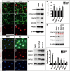
Wnt/β-catenin signaling is necessary and sufficient to regulate the expression of VEC markers in LECs. (AD) Primary human LECs were cultured in the presence or absence of OSS for 48 h. Subsequently, cells were analyzed by IHC for the indicated markers (A,B), Western blot (C), or quantitative PCR (qPCR) (D). (A,B) IHC revealed the up-regulation of FOXC2 and β-catenin expression by OSS. (C) Western blot revealed an increase in the expression levels of total and active β-catenin. Valve-expressed transcription factors GATA2 and FOXC2 are also increased. PROX1 levels are not obviously changed. (D) qPCR validated the up-regulation of FOXC2. Additionally, target genes of the Wnt/β-catenin signaling pathway—Axin2, Cyclin-D1, and c-Jun—are increased. (E) LECs were cultured under static or OSS conditions in the presence or absence of 25 µM iCRT3, an antagonist of Wnt/β-catenin signaling, for 48 h. Western blot revealed a modest down-regulation of FOXC2, GATA2, and PROX1 expression by iCRT3 under static conditions. In contrast, iCRT3 dramatically inhibited the OSS-mediated up-regulation of FOXC2 and GATA2 expressions. The numbers in red indicate the relative expression of FOXC2 as measured densitometrically. (FI) Primary human LECs were cultured in the presence of 0.5 µM Bio, an agonist of the Wnt/β-catenin signaling pathway, for 12 h. Subsequently, cells were analyzed by IHC for the indicated markers (F,G), Western blot (H), or qPCR (I). The results show that Bio enhances the expression of valve markers FOXC2, GATA2, PROX1, and CX37. Bars: A, B, F, G, 100 µm. n = 3 for each experiment. (*) P < 0.05; (**) P < 0.01; (***) P < 0.001.
Figure 6.
Figure 6.
β-Catenin directly associates with the regulatory elements of FOXC2 and PROX1. (A) TCF/LEF family transcription factors associate with the indicated motif. β-Catenin recognizes and binds this consensus sequence via TCF/LEF when Wnt/β-catenin signaling is active. (B) Genomic alignment was performed using the regulatory elements of the genes encoding FOXC2 from various mammals. Nucleotides in red are highly conserved. A highly conserved TCF/LEF-binding site is observed in the regulatory elements of FOXC2 (enclosed within the red box). (C) The TCF/LEF-binding site (red arrow) is located ∼3.5 kb upstream of the TSS of FOXC2. Based on ChIP-seq data curated by The ENCODE Project Consortium (2012), the TCF/LEF-binding site is located in a region (blue box) that is enriched for H3K4me3 and H3K27ac histone modifications that are associated with transcriptionally active genes. (D) Primary human LECs were cultured in the presence of 0.5 µM DMSO or Bio for 12 h. Subsequently, cells were harvested, and ChIP was performed using IgG or an antibody specific to β-catenin. PCR was performed using primers flanking the TCF/LEF-binding site in the −3.5-kb location. As a negative control, primers flanking a TCF/LEF-binding site that is located at a more upstream location (−5.5 kb) were used. Gel and qPCR results show that β-catenin associates with the −3.5-kb site but not the −5.5-kb site in Bio-treated LECs. (E) β-Catenin associates with the TCF/LEF-binding sites in the regulatory elements of PROX1 at the indicated locations. This interaction is significantly enhanced by Bio. (D,E) n = 3. (*) P < 0.05; (**) P < 0.01.
Figure 7.
Figure 7.
FOXC2 compensates for the loss of β-catenin to regulate lymphatic vessel patterning. (AE) Primary human LECs were infected with control or FOXC2-expressing retroviral particles. Scratch assay was performed 24 h later in the presence or absence of 25 µM iCRT3. The space between the red dotted lines indicates the open scratch wound. (E) The wound size was measured at various time points and plotted. iCRT3 significantly inhibits the ability of control LECs to “heal” the scratch wound. FOXC2-overexpressing cells are able to significantly overcome iCRT3-induced inhibition. (FI) The lymphatic vessels of the dorsal skin of E16.5 control (F), Lyve1-Cre;Ctnnb1LOF (G), Lyve1-Cre;Ctnnb1LOF;FOXC2GOF (H), and Lyve1-Cre;FOXC2GOF (I) embryos were analyzed by IHC for VEGFR3. (J) The diameter of the vessels and the distance between the tip cells of the opposing fronts were quantified (Supplemental Fig. 7E–H) and plotted. In comparison with control embryos, the lymphatic vessels of Lyve1-Cre;Cttnb1LOF embryos are significantly dilated. The distance between the migrating fronts is also significantly increased, indicating lymphatic vascular hypoplasia. Ectopic expression of FOXC2 significantly rescues these defects. (KN) Coimmunohistochemistry for the indicated markers revealed the presence of α-SMA+ mural cells on the VEGFR3+ lymphatic vessels of Lyve1-Cre;Cttnb1LOF embryos (shown in L). (M) This defect is fully rescued by ectopic expression of FOXC2. Lower magnification pictures of FI and KN are presented in Supplemental Figure 7. (OR) The mesenteric lymphatic vessels of E17.5 embryos were analyzed by IHC for the indicated markers. Dilation of lymphatic vessels and the abnormal recruitment of α-SMA+ mural cells caused by the loss of β-catenin were rescued by FOXC2 overexpression. LVs are seen in control (arrow) but not in any of the mutant embryos. (S) Model for the relationship between OSS, β-catenin, FOXC2, and PROX1 during lymphatic vascular development. We did not exclude the role of yet to be identified Wnt ligands that function independently or in cooperation with OSS to activate β-catenin. Bars, 100 µm. (AE) n = 3. (FR) n = 4. (*) P < 0.05; (**) P < 0.01; (***) P < 0.001.

References

    1. Al Alam D, Green M, Tabatabai Irani R, Parsa S, Danopoulos S, Sala FG, Branch J, El Agha E, Tiozzo C, Voswinckel R, et al. 2011. Contrasting expression of canonical Wnt signaling reporters TOPGAL, BATGAL and Axin2(LacZ) during murine lung development and repair. PLoS One 6: e23139. - PMC - PubMed
    1. Alfieri CM, Cheek J, Chakraborty S, Yutzey KE. 2010. Wnt signaling in heart valve development and osteogenic gene induction. Dev Biol 338: 127–135. - PMC - PubMed
    1. Bazigou E, Xie S, Chen C, Weston A, Miura N, Sorokin L, Adams R, Muro AF, Sheppard D, Makinen T. 2009. Integrin-α9 is required for fibronectin matrix assembly during lymphatic valve morphogenesis. Dev Cell 17: 175–186. - PMC - PubMed
    1. Berger J, Berger S, Tuoc TC, D'Amelio M, Cecconi F, Gorski JA, Jones KR, Gruss P, Stoykova A. 2007. Conditional activation of Pax6 in the developing cortex of transgenic mice causes progenitor apoptosis. Development 134: 1311–1322. - PubMed
    1. Bertozzi CC, Schmaier AA, Mericko P, Hess PR, Zou Z, Chen M, Chen CY, Xu B, Lu MM, Zhou D, et al. 2010. Platelets regulate lymphatic vascular development through CLEC-2–SLP-76 signaling. Blood 116: 661–670. - PMC - PubMed

Publication types

Substances