Review
. 2016:2016:8562805.
doi: 10.1155/2016/8562805. Epub 2016 May 25.

Neuroinvasion and Inflammation in Viral Central Nervous System Infections

Affiliations
Review

Neuroinvasion and Inflammation in Viral Central Nervous System Infections

Tobias Dahm et al. Mediators Inflamm. 2016.

Abstract

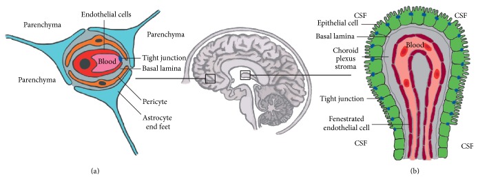
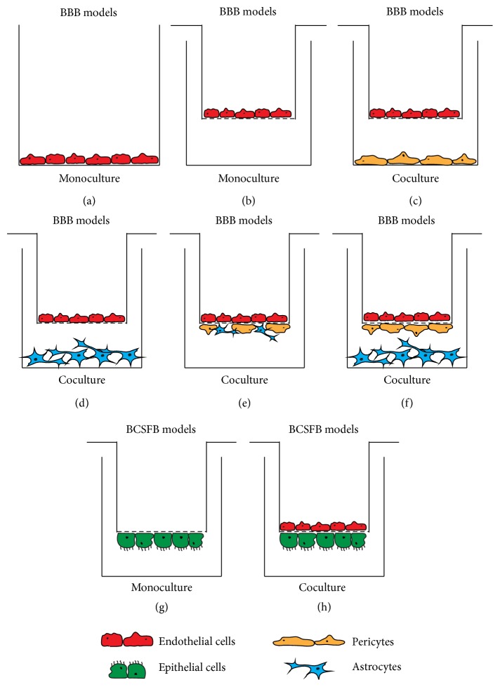
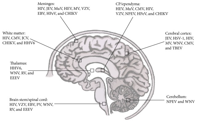
Neurotropic viruses can cause devastating central nervous system (CNS) infections, especially in young children and the elderly. The blood-brain barrier (BBB) and the blood-cerebrospinal fluid barrier (BCSFB) have been described as relevant sites of entry for specific viruses as well as for leukocytes, which are recruited during the proinflammatory response in the course of CNS infection. In this review, we illustrate examples of established brain barrier models, in which the specific reaction patterns of different viral families can be analyzed. Furthermore, we highlight the pathogen specific array of cytokines and chemokines involved in immunological responses in viral CNS infections. We discuss in detail the link between specific cytokines and chemokines and leukocyte migration profiles. The thorough understanding of the complex and interrelated inflammatory mechanisms as well as identifying universal mediators promoting CNS inflammation is essential for the development of new diagnostic and treatment strategies.


Figures

Figure 1
Figure 1
Barriers of the brain. (a) The BBB found between the lumen of the cerebral blood vessels and the brain parenchyma is composed of capillary endothelial cells, astrocytes, and pericytes and obtains its characteristic physical barrier function via linking the endothelial cells through luminal tight junctions. (b) The BCSFB located between the CSF and the fenestrated blood vessels of the choroid plexus. This selective barrier contains polar choroid plexus epithelial cells joined to one another through tight junctions found on the apical side.
Figure 2
Figure 2
Schematic representation of various in vitro BBB and BCSFB models. The BBB models are built up around endothelial cells, whereas the BCSFB models rely on epithelial cell culture. The setup can be a monoculture: endothelial or epithelial cells only (a, b, and g); coculture: endothelial cells with epithelial cells (h) or endothelial cells with pericytes or astrocytes (c and d); and triple-culture: endothelial in combination with astrocytes and pericytes (e and f). These systems can be set up in a “contact” (e, f, and h) or “noncontact” (c and d) manner (“contact”: different cell types have physical contact with each other; “noncontact”: different cell types were not able to physically interact with each other). The monoculture models represented in (a) and (b) can also be used with epithelial cells instead of endothelial cells. The addition of chemical mediators (cytokines and/or chemokines) and virus or immune cells (T-, B-cells, and PMNs, etc.) is possible in both the filter insert and/or well.
Figure 3
Figure 3
Relevant encephalitis and meningitis causing viruses in humans allocated to the brain region they may affect. CHIKV, chikungunya virus; CMV, cytomegalovirus; EBV, Epstein Barr virus; EEEV, eastern equine encephalitis virus; HHV-6, human herpes virus-6; HIV, human immunodeficiency virus; HPeV, human parechovirus; HSV-1, herpes simplex virus-1; JCV, John Cunningham virus; JEV, Japanese encephalitis virus; MV, measles virus; MuV, mumps virus; NPEV, nonpolio enterovirus; PV, poliovirus; RV, rabies virus; TBEV, tick-borne encephalitis virus; WNV, West Nile virus; VZV, varicella zoster virus.

Similar articles

Cited by

References

    1. Swanson P. A., II, McGavern D. B. Viral diseases of the central nervous system. Current Opinion in Virology. 2015;11:44–54. doi: 10.1016/j.coviro.2014.12.009. - DOI - PMC - PubMed
    1. Michos A. G., Syriopoulou V. P., Hadjichristodoulou C., et al. Aseptic meningitis in children: analysis of 506 cases. PloS ONE. 2007;2(7, article e674) - PMC - PubMed
    1. Rotbart H. A. Viral meningitis. Seminars in Neurology. 2000;20(3):277–292. doi: 10.1055/s-2000-9427. - DOI - PubMed
    1. Rantakallio P., Leskinen M., von Wendt L. Incidence and prognosis of central nervous system infections in a birth cohort of 12,000 children. Scandinavian Journal of Infectious Diseases. 1986;18(4):287–294. doi: 10.3109/00365548609032339. - DOI - PubMed
    1. Kivisäkk P., Mahad D. J., Callahan M. K., et al. Human cerebrospinal fluid central memory CD4+ T cells: evidence for trafficking through choroid plexus and meninges via P-selectin. Proceedings of the National Academy of Sciences of the United States of America. 2003;100(14):8389–8394. doi: 10.1073/pnas.1433000100. - DOI - PMC - PubMed

MeSH terms

LinkOut - more resources