. 2016:2016:1473918.
doi: 10.1155/2016/1473918. Epub 2016 May 29.

Correlation Assessment between Three-Dimensional Facial Soft Tissue Scan and Lateral Cephalometric Radiography in Orthodontic Diagnosis

Affiliations

Correlation Assessment between Three-Dimensional Facial Soft Tissue Scan and Lateral Cephalometric Radiography in Orthodontic Diagnosis

Piero Antonio Zecca et al. Int J Dent. 2016.

Abstract

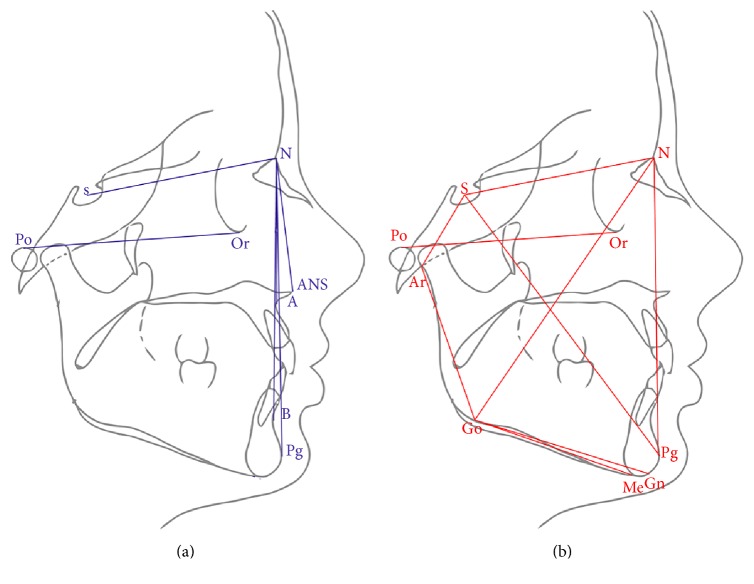
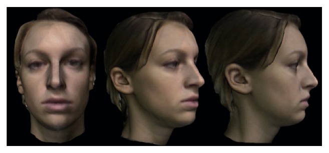
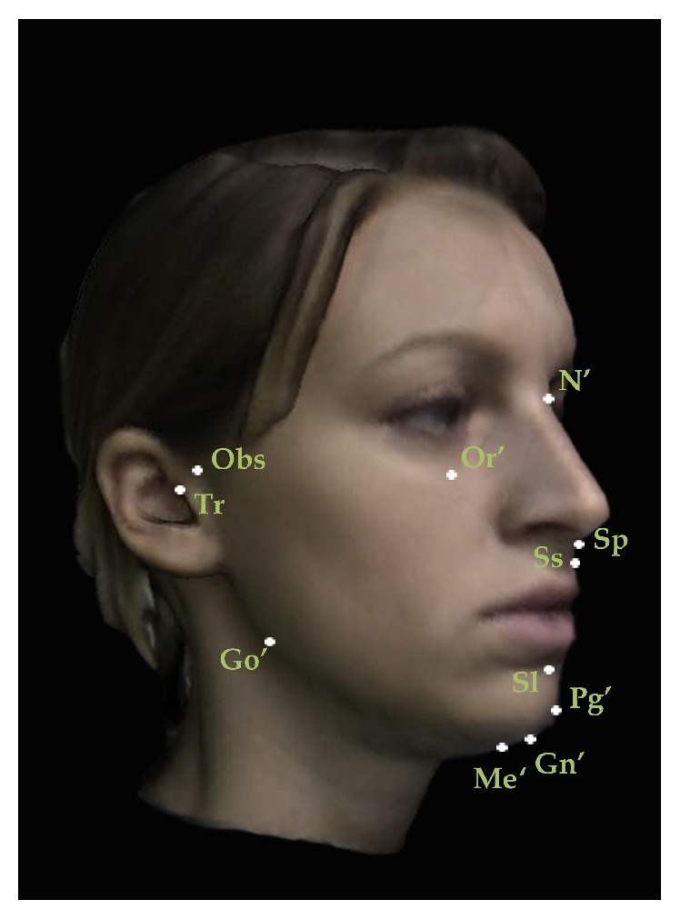
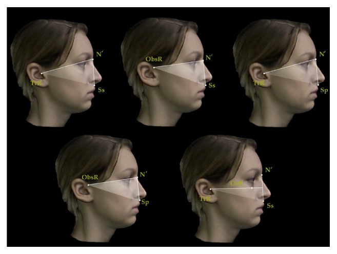
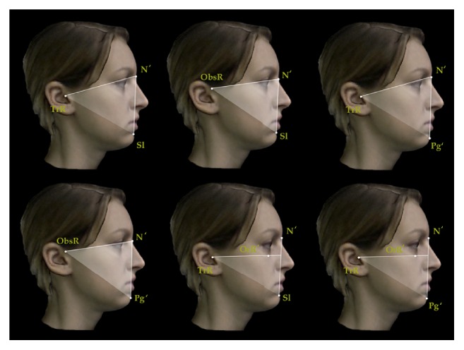
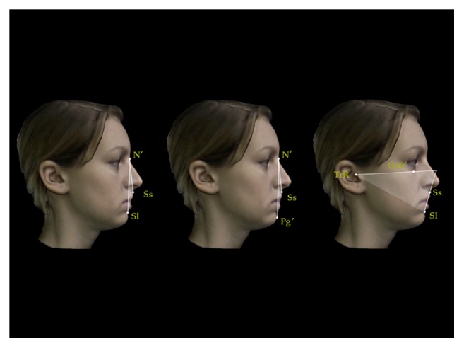
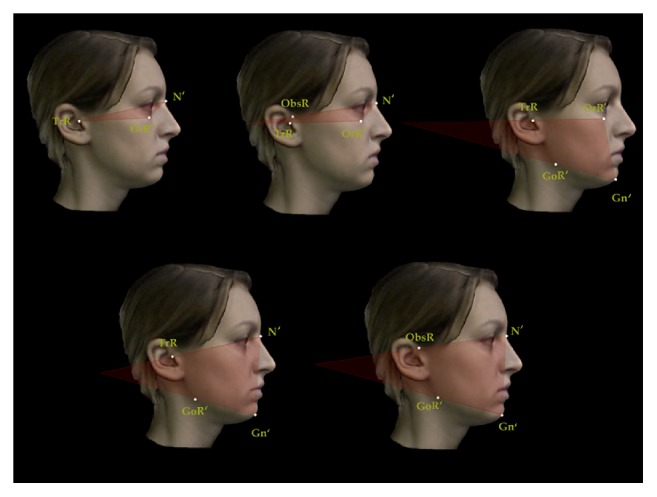
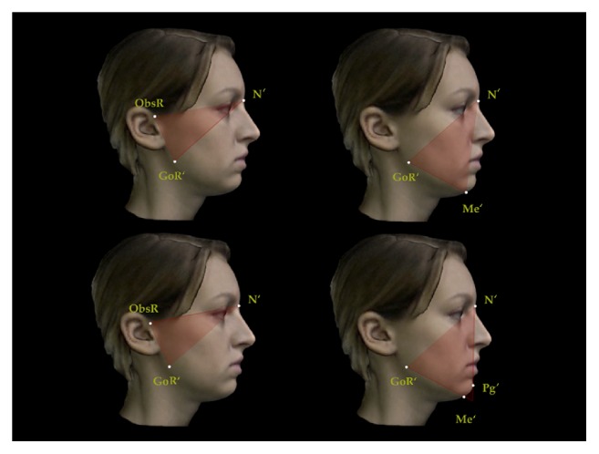
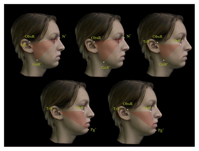
Purpose. The aim of the present prospective study was to investigate correlations between 3D facial soft tissue scan and lateral cephalometric radiography measurements. Materials and Methods. The study sample comprised 312 subjects of Caucasian ethnic origin. Exclusion criteria were all the craniofacial anomalies, noticeable asymmetries, and previous or current orthodontic treatment. A cephalometric analysis was developed employing 11 soft tissue landmarks and 14 sagittal and 14 vertical angular measurements corresponding to skeletal cephalometric variables. Cephalometric analyses on lateral cephalometric radiographies were performed for all subjects. The measurements were analysed in terms of their reliability and gender-age specific differences. Then, the soft tissue values were analysed for any correlations with lateral cephalometric radiography variables using Pearson correlation coefficient analysis. Results. Low, medium, and high correlations were found for sagittal and vertical measurements. Sagittal measurements seemed to be more reliable in providing a soft tissue diagnosis than vertical measurements. Conclusions. Sagittal parameters seemed to be more reliable in providing a soft tissue diagnosis similar to lateral cephalometric radiography. Vertical soft tissue measurements meanwhile showed a little less correlation with the corresponding cephalometric values perhaps due to the low reproducibility of cranial base and mandibular landmarks.


Figures

Figure 1
Figure 1
2D tracings and cephalometric analysis performed with lateral cephalometric radiography. (a) Sagittal measurements and (b) vertical measurements.
Figure 2
Figure 2
Facial soft tissue scan. Frontal, prospective, and right lateral 3D views of facial soft tissue scan of female patient.
Figure 3
Figure 3
Set of reproducible landmarks employed to perform sagittal and vertical soft tissue 3D measurements: N′ Nasion; Obs Otobasion; Tr Tragus; Or′ Orbitale; Sp Spinal; Ss Subspinal; Go′ Gonion; Sl Sublabial; Pg′ Pogonion; Me′ Menton; Gn′ Gnation.
Figure 4
Figure 4
Sagittal angular measurements for 3D facial soft tissue. R: right. Maxillary sagittal measurements.
Figure 5
Figure 5
Sagittal angular measurements for 3D facial soft tissue. R: right. Mandibular sagittal measurements.
Figure 6
Figure 6
Sagittal angular measurements for 3D facial soft tissue. R: right. Maxillomandibular sagittal measurements.
Figure 7
Figure 7
Vertical angular measurements for 3D facial soft tissue. R: right. Part 1.
Figure 8
Figure 8
Vertical angular measurements for 3D facial soft tissue. R: right. Part 2.
Figure 9
Figure 9
Vertical angular measurements for 3D facial soft tissue. R: right. Part 3.

References

    1. Nijkamp P. G., Habets L. L. M. H., Aartman I. H. A., Zentner A. The influence of cephalometrics on orthodontic treatment planning. European Journal of Orthodontics. 2008;30(6):630–635. doi: 10.1093/ejo/cjn059. - DOI - PubMed
    1. Kilic N., Catal G., Kiki A., Oktay H. Soft tissue profile changes following maxillary protraction in Class III subjects. European Journal of Orthodontics. 2010;32(4):419–424. doi: 10.1093/ejo/cjp119. - DOI - PubMed
    1. Devereux L., Moles D., Cunningham S. J., McKnight M. How important are lateral cephalometric radiographs in orthodontic treatment planning? American Journal of Orthodontics and Dentofacial Orthopedics. 2011;139(2):e175–e181. doi: 10.1016/j.ajodo.2010.09.021. - DOI - PubMed
    1. Arnett G. W., Bergman R. T. Facial keys to orthodontic diagnosis and treatment planning. Part I. American Journal of Orthodontics and Dentofacial Orthopedics. 1993;103(4):299–312. doi: 10.1016/0889-5406(93)70010-L. - DOI - PubMed
    1. Arnett G. W., Bergman R. T. Facial keys to orthodontic diagnosis and treatment planning. Part II. American Journal of Orthodontics and Dentofacial Orthopedics. 1993;103(5):395–411. doi: 10.1016/s0889-5406(05)81791-3. - DOI - PubMed