. 2016:2016:1739760.
doi: 10.1155/2016/1739760. Epub 2016 May 24.

Hataedock Treatment Has Preventive Therapeutic Effects in Atopic Dermatitis-Induced NC/Nga Mice under High-Fat Diet Conditions

Affiliations

Hataedock Treatment Has Preventive Therapeutic Effects in Atopic Dermatitis-Induced NC/Nga Mice under High-Fat Diet Conditions

Ho-Yeol Cha et al. Evid Based Complement Alternat Med. 2016.

Abstract

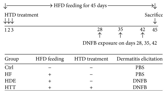
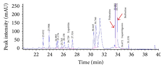
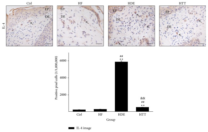
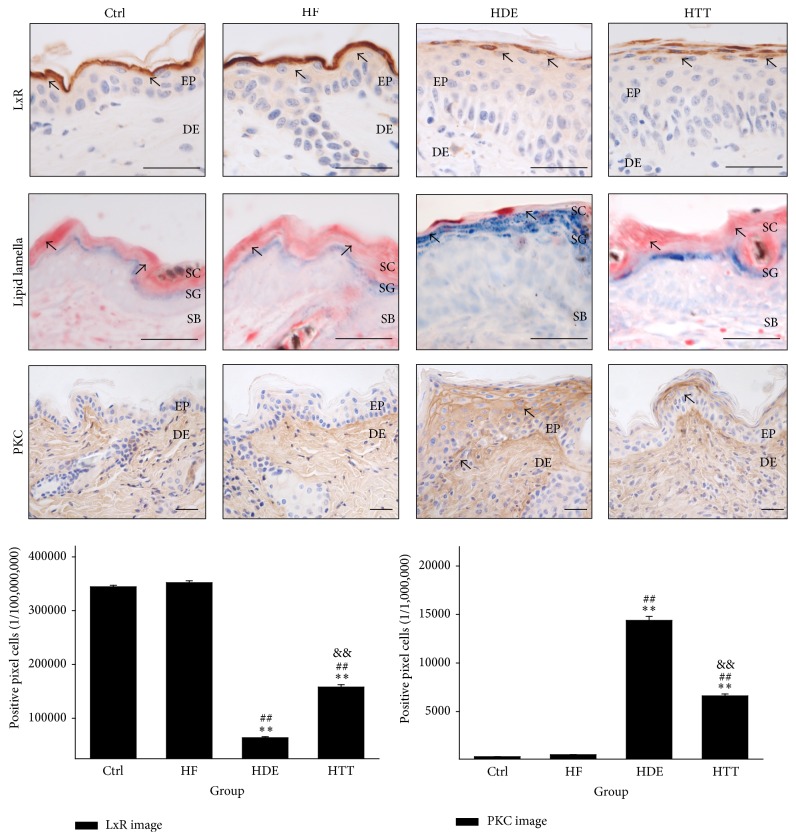
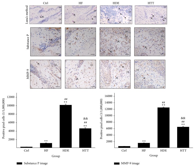
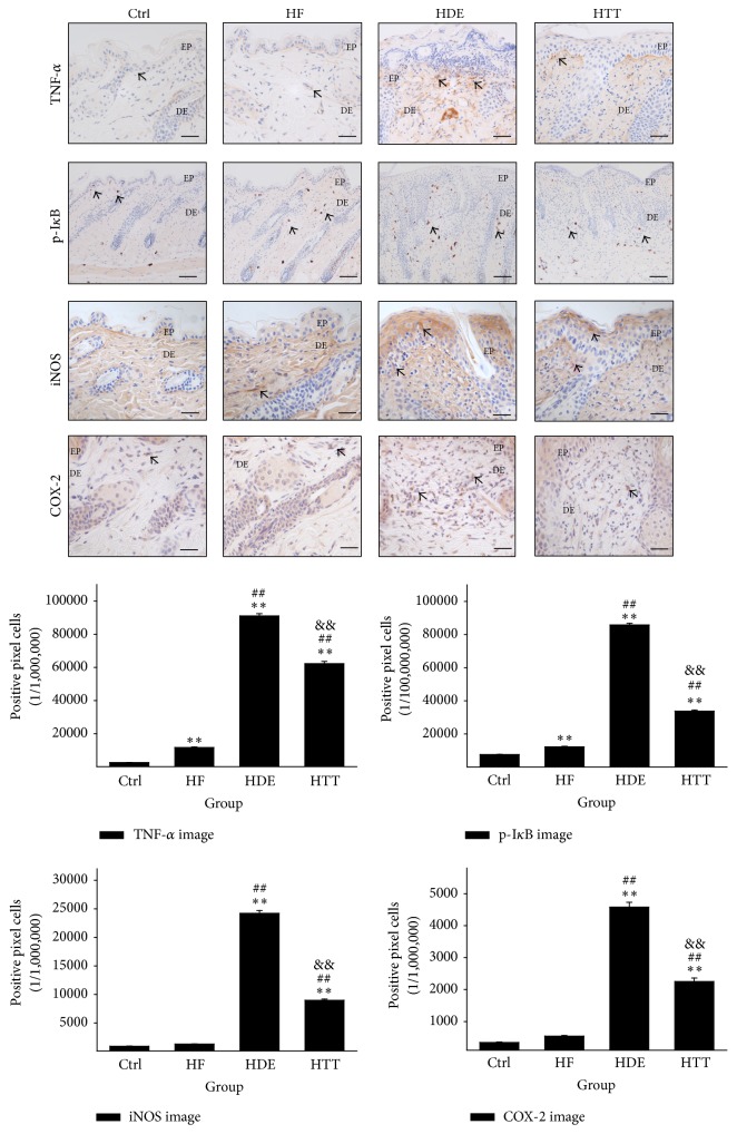
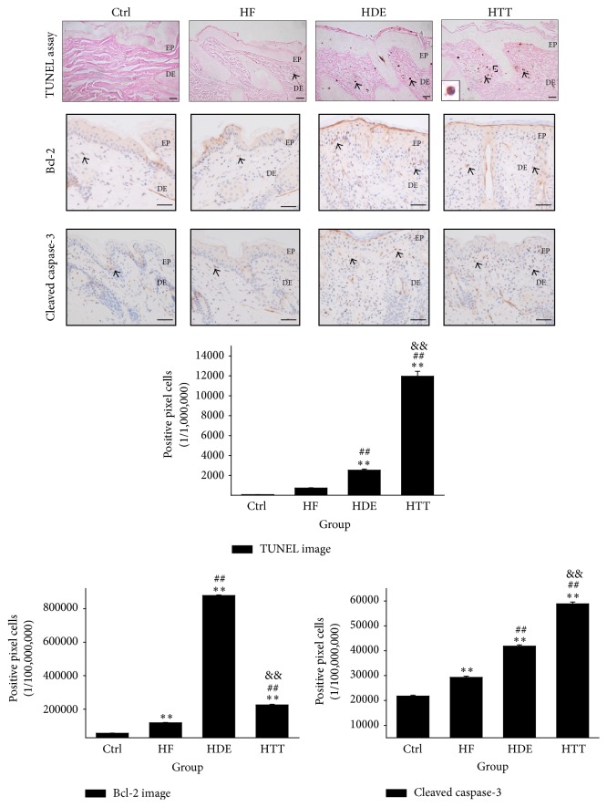
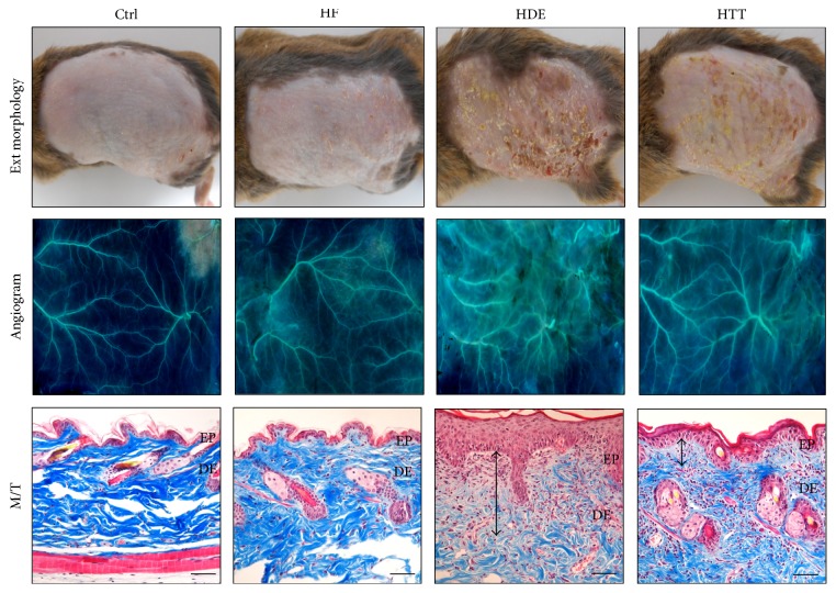
This study investigated the preventive therapeutic effects of Hataedock (HTD) treatment on inflammatory regulation and skin protection in AD-induced NC/Nga mice under high-fat diet conditions. Before inducing AD, the extract of Coptidis Rhizoma and Glycyrrhiza uralensis was administered orally to the 3-week-old mice. After that, AD-like skin lesions were induced by applying DNFB. All groups except the control group were fed a high-fat diet freely. We identified the effects of HTD on morphological changes, cytokine release and the induction of apoptosis through histochemistry, immunohistochemistry, and TUNEL assay. HTD downregulated the levels of IL-4 and PKC but increased the levels of LXR. HTD also suppressed the mast cell degranulation and release of MMP-9, Substance P. The levels of TNF-α, p-IκB, iNOS, and COX-2 were also decreased. The upregulation of inflammatory cell's apoptosis is confirmed by our results as increase of apoptotic body and cleaved caspase-3 and decrease of Bcl-2. HTD also reduced edema, angiogenesis, and skin lesion inflammation. Our results indicate HTD suppresses various inflammatory response on AD-induced mice with obesity through the regulation of Th2 differentiation and the protection of lipid barrier. Therefore, HTD could be used as an alternative and preventive therapeutic approach in the management of AD.


Figures

Figure 1
Figure 1
Experimental design. Before inducing AD, the extract of Coptidis Rhizoma and Glycyrrhiza uralensis, which is traditionally used in HTD treatment, was administered orally to the HTT group on days 1, 2, and 3. Mice were challenged by DNFB on days 28, 35, and 42. During the experimental period, all groups except the Ctrl group were fed a high-fat diet freely. Ctrl: normal feeding, HF: high-fat diet, HDE: high-fat diet and untreated AD-induced, HTT: high fat diet and Hataedock treated AD-induced, HFD: high-fat diet, HTD: Hataedock, PBS: phosphate-buffered saline, and DNFB: dinitrofluorobenzene.
Figure 2
Figure 2
The HPLC analysis of the extract of Coptidis Rhizoma and Glycyrrhiza uralensis. Palmatine was detected at approximately 33.001 minutes, Berberine was detected at approximately 33.775 minutes, Liquritin was detected at approximately 26.729 minutes, and Liquiritigenin was detected at approximately 34.672 minutes. HPLC: High-performance liquid chromatography.
Figure 3
Figure 3
The regulation of Th2 differentiation. IL-4-positive reaction (arrow indicates dark brown) decreased in the HTT group compared with the HDE group (IL-4 immunohistochemistry; bar size, 50 μm). Data of IL-4 image analysis was also showing the same result (p < 0.01). Ctrl: normal feeding, HF: high-fat diet, HDE: high-fat diet and untreated AD-induced, HTT: high fat diet and Hataedock treated AD-induced, EP: epidermis, and DE: dermis. ∗∗ p < 0.01, compared with the Ctrl group; ## p < 0.01, compared with the HF group; && p < 0.01, compared with the HDE group.
Figure 4
Figure 4
The maintenance of lipid barrier in epidermis. The LXR-positive reaction (arrow indicates dark brown) in HDE remarkably decreased but was maintained in HTT (LXR immunohistochemistry; bar size, 50 μm). The intercellular lipid lamellae (arrow indicates reddish line) of the stratum corneum disappeared in HDE but appeared in HTT (oil red O; bar size, 50 μm). The PKC-positive reaction (arrow indicates dark brown) in HTT remarkably decreased (PKC immunohistochemistry; bar size, 50 μm). Data of LXR and PKC image analysis was also showing the same result (p < 0.01). Ctrl: normal feeding, HF: high-fat diet, HDE: high-fat diet and untreated AD-induced, HTT: high fat diet and Hataedock treated AD-induced, EP: epidermis, DE: dermis, SC: stratum corneum, SG: stratum granulosum, SB: stratum basale, LXR: liver X receptor, and PKC: protein kinase C. ∗∗ p < 0.01, compared with the Ctrl group; ## p < 0.01, compared with the HF group; && p < 0.01, compared with the HDE group.
Figure 5
Figure 5
The regulation of mast cells activation. The distribution of degranulated mast cell (vacant arrow) in dermal papillae was increased in the HDE group but decreased in the HTT group (Luna's method; bar size, 50 μm). The Substance P positive reaction (arrow indicates dark brown) in HTT significantly decreased (Substance P immunohistochemistry; bar size, 50 μm). The MMP-9 positive reaction (arrow indicates dark brown) in HTT remarkably decreased (MMP-9 immunohistochemistry; bar size, 50 μm). Data of Substance P and MMP-9 image analysis was also showing the same result (p < 0.01). Ctrl: normal feeding, HF: high-fat diet, HDE: high-fat diet and untreated AD-induced, HTT: high fat diet and Hataedock treated AD-induced, EP: epidermis, DE: dermis, and MMP-9: matrix metalloproteinases-9. ∗∗ p < 0.01, compared with the Ctrl group; ## p < 0.01, compared with the HF group; && p < 0.01, compared with the HDE group.
Figure 6
Figure 6
Downregulation of inflammation. In HTT group, the excessive inflammation condition such as increase of TNF-α, p-IκB, iNOS, and COX-2 was ameliorated by HTD treatment. These positive reactions (arrow indicates dark brown) were remarkably decreased compared with those of the HDE group (immunohistochemistry; bar size, 50 μm, only p-IκB bar size 100 μm). Data of TNF-α, p-IκB, iNOS, and COX-2 image analysis was also showing the same result (p < 0.01). Ctrl: normal feeding, HF: high-fat diet, HDE: high-fat diet and untreated AD-induced, HTT: high fat diet and Hataedock treated AD-induced, EP: epidermis, DE: dermis. ∗∗ p < 0.01, compared with the Ctrl group; ## p < 0.01, compared with the HF group; && p < 0.01, compared with the HDE group.
Figure 7
Figure 7
Upregulation of apoptosis. Upregulation of apoptosis in dermatitis induced by HTD treatment. The apoptotic body (arrow indicates dark brown) in the HTT group was remarkably increased compared to the HDE group (TUNEL assay; square box, enlarged DNA fragmentation of nucleus with TUNEL positive reaction; bar size, 50 μm). The Bcl-2 positive reaction (arrow indicates dark brown) in HTT group was remarkably decreased compared with those of the HDE group (immunohistochemistry; bar size, 50 μm). The cleaved caspase-3 positive reaction (arrow indicates dark brown) in HTT group was increased compared with those of the HDE group (immunohistochemistry; bar size, 50 μm). Data of TUNEL, Bcl-2, and cleaved caspase-3 image analysis was also showing the same result (p < 0.01). Ctrl: normal feeding, HF: high-fat diet, HDE: high-fat diet and untreated AD-induced, HTT: high fat diet and Hataedock treated AD-induced, EP: epidermis, and DE: dermis. ∗∗ p < 0.01, compared with the Ctrl group; ## p < 0.01, compared with the HF group; && p < 0.01, compared with the HDE group.
Figure 8
Figure 8
The mitigative effect of HTD treatment for dermatitis. The skin damage as eczema was mitigated in HTT group. The angiogenesis was increased in HDE group but decreased in HTT group (×4). The histological features of AD such as vacuolation of keratinocytes, hyperplasia, edema (up-down arrow), infiltration of inflammatory cells, and increase of capillary were increased in HDE group but decreased in HTT group (bar size, 100 μm). Ctrl: normal feeding, HF: high-fat diet, HDE: high-fat diet and untreated AD-induced, HTT: high fat diet and Hataedock treated AD-induced, EP: epidermis, DE: dermis, and M/T: Masson trichrome method.

Similar articles

Cited by

References

    1. Behniafard N., Gharagozlou M., Farhadi E., et al. TNF-alpha single nucleotide polymorphisms in atopic dermatitis. European Cytokine Network. 2012;23(4):163–165. doi: 10.1684/ecn.2012.0323. - DOI - PubMed
    1. Kawakami T., Ando T., Kimura M., Wilson B. S., Kawakami Y. Mast cells in atopic dermatitis. Current Opinion in Immunology. 2009;21(6):666–678. doi: 10.1016/j.coi.2009.09.006. - DOI - PMC - PubMed
    1. Barnes K. C. An update on the genetics of atopic dermatitis: scratching the surface in 2009. Journal of Allergy and Clinical Immunology. 2010;125(1–3):16.e11–29.e11. doi: 10.1016/j.jaci.2009.11.008. - DOI - PMC - PubMed
    1. Danby S., Cork M. J. A new understanding of atopic dermatitis: the role of epidermal barrier dysfunction and subclinical inflammation. Journal of Clinical Dermatology. 2012;1:33–46.
    1. Elias P. M., Schmuth M. Abnormal skin barrier in the etiopathogenesis of atopic dermatitis. Current Opinion in Allergy and Clinical Immunology. 2009;9(5):437–446. doi: 10.1097/ACI.0b013e32832e7d36. - DOI - PMC - PubMed