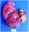
Recurrent primary mediastinal liposarcoma: A case report
- PMID: 27313694
- PMCID: PMC4888245
- DOI: 10.3892/ol.2016.4453
Recurrent primary mediastinal liposarcoma: A case report
Abstract
Primary mediastinal liposarcomas are extremely rare. The current study reports the case of a 63-year-old man presenting with a primary liposarcoma arising from the posterior mediastinum. The patient reported a 6-month history of chest pain with increasing dyspnea for 2 months. Enhanced computed tomography revealed a 10×16-cm mass in the posterior mediastinum. Other physical examinations were normal. Radical resection was performed under the agreement of patient. Subsequent pathological analysis indicated a liposarcoma. The patient recovered and was successfully discharged. However, at a follow-up examination 12 months after surgery, recurrence was identified in the anterior mediastinum. Therefore, the patient underwent surgery. The postoperative course was uneventful, however, there was evidence of disease recurrence 2 years after the second surgery. The patient refused any treatment and succumbed after 3 months.
Keywords: mediastinal liposarcoma; recurrent; surgery.
Figures


References
-
- Punpale A, Pramesh CS, Jambhekar N, Mistry RC. Giant mediastinal liposarcoma: A case report. Ann Thorac Cardiovasc Surg. 2006;12:425–427. - PubMed
-
- Wick MR. The mediastinum. In: Sternberg SS, Antonioli DA, editors. Diagnostic Surgical Pathology. 3rd. 1 and 2. Lippincott Williams and Wilkins; Philadelphia, PA: 1999. pp. 1147–1208.
LinkOut - more resources
Full Text Sources
Other Literature Sources
