

Liposome-mediated transfection of wild-type P53 DNA into human prostate cancer cells is improved by low-frequency ultrasound combined with microbubbles
- PMID: 27313702
- PMCID: PMC4888269
- DOI: 10.3892/ol.2016.4477
Liposome-mediated transfection of wild-type P53 DNA into human prostate cancer cells is improved by low-frequency ultrasound combined with microbubbles
Abstract
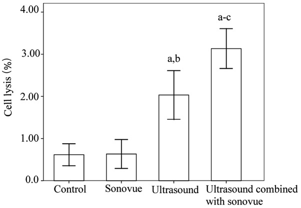
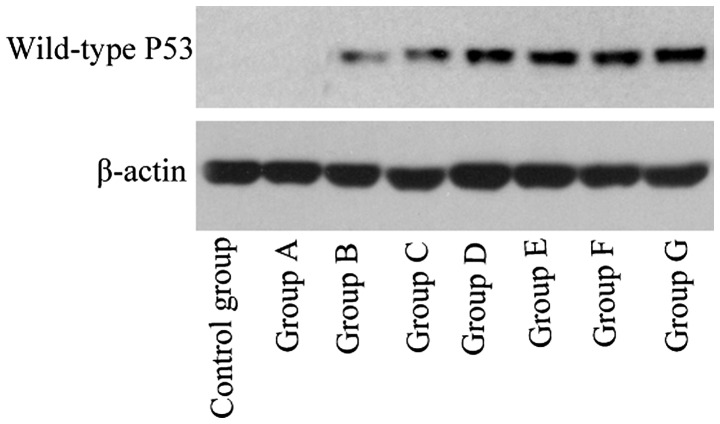
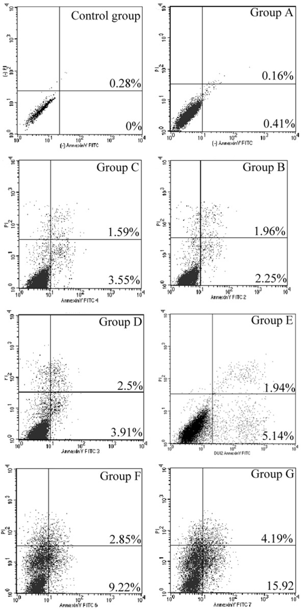
Prostate cancer is a common type of cancer in elderly men. The aim of the present study was to evaluate the effects of ultrasound exposure in combination with SonoVue microbubbles on liposome-mediated transfection of wild-type P53 genes into human prostate cancer cells. PC-3 human prostate cancer cells were exposed to ultrasound; duty cycle was controlled at 20% (2 sec on, 8 sec off) for 5 min with and without SonoVue microbubble echo-contrast agent using a digital sonifier (frequency, 21 kHz; intensity, 46 mW/cm2). The cells were divided into eight groups, as follows: Group A (SonoVue + wild-type P53), group B (ultrasound + wild-type P53), group C (SonoVue + ultrasound + wild-type P53), group D (liposome + wild-type P53), group E (liposome + SonoVue + wild-type P53), group F (liposome + wild-type P53 + ultrasound), group G (liposome + wild-type P53 + ultrasound + SonoVue) and the control group (wild-type P53). Following treatment, a hemocytometer was used to measure cell lysis, reverse transcription-quantitative polymerase chain reaction and western blotting were performed to detect P53 gene transfection efficiency, Cell Counting Kit-8 was employed to reveal cell proliferation and Annexin V/propidium iodide staining was used to determine cell apoptosis. Cell lysis was minimal in each group. Wild-type P53 gene and protein expression were significantly increased in the PC-3 cells in group G compared with the control and all other groups (P<0.01). Cell proliferation was significantly suppressed in group G compared with the control group and all other groups (P<0.01). Cell apoptosis levels in group G were significantly improved compared with the control group and all other groups (P<0.01). Thus, the results of the present study indicate that the use of low-frequency and low-energy ultrasound in combination with SonoVue microbubbles may be a potent physical method for increasing liposome gene delivery efficiency.
Keywords: gene therapy; liposome; low-energy ultrasound; low-frequency ultrasound; microbubble; wild-type P53.
Figures

References
-
- Lecornet E, Ahmed HU, Moore C, Emberton M. Focal therapy for prostate cancer: A potential strategy to address the problem of overtreatment. Arch Esp Urol. 2010;63:845–852. - PubMed
LinkOut - more resources
Full Text Sources
Other Literature Sources
Research Materials
Miscellaneous