

Vitamin D3 Suppresses Class II Invariant Chain Peptide Expression on Activated B-Lymphocytes: A Plausible Mechanism for Downregulation of Acute Inflammatory Conditions
- PMID: 27313879
- PMCID: PMC4904097
- DOI: 10.1155/2016/4280876
Vitamin D3 Suppresses Class II Invariant Chain Peptide Expression on Activated B-Lymphocytes: A Plausible Mechanism for Downregulation of Acute Inflammatory Conditions
Abstract
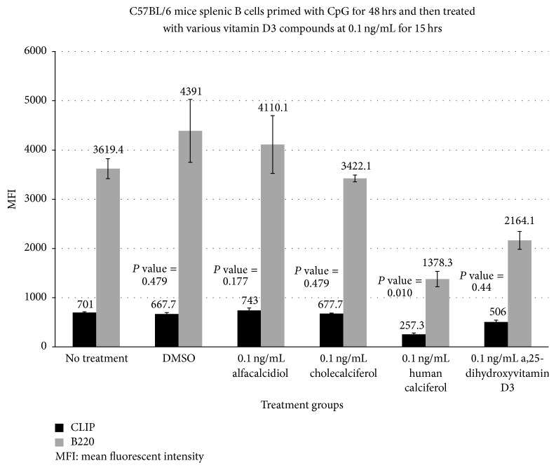
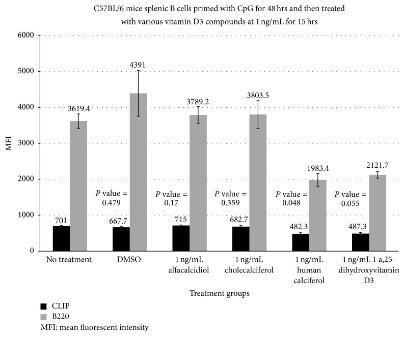
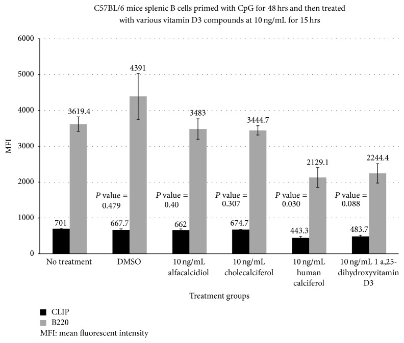
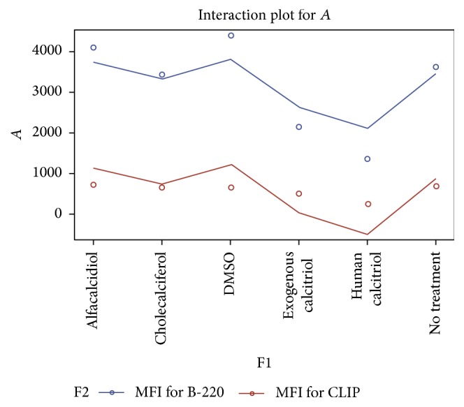
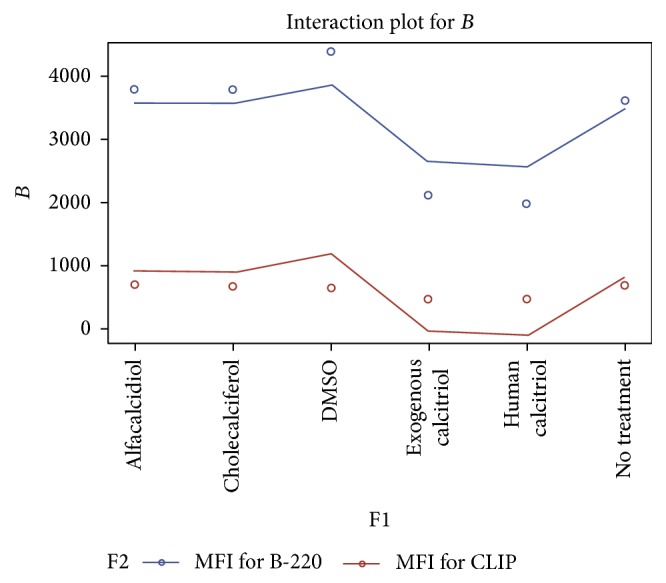
Class II invariant chain peptide (CLIP) expression has been demonstrated to play a pivotal role in the regulation of B cell function after nonspecific polyclonal expansion. Several studies have shown vitamin D3 helps regulate the immune response. We hypothesized that activated vitamin D3 suppresses CLIP expression on activated B-cells after nonspecific activation or priming of C57BL/6 mice with CpG. This study showed activated vitamin D3 actively reduced CLIP expression and decreased the number of CLIP(+) B-lymphocytes in a dose and formulation dependent fashion. Flow cytometry was used to analyze changes in mean fluorescent intensity (MFI) based on changes in concentration of CLIP on activated B-lymphocytes after treatment with the various formulations of vitamin D3. The human formulation of activated vitamin D (calcitriol) had the most dramatic reduction in CLIP density at an MFI of 257.3 [baseline of 701.1 (P value = 0.01)]. Cholecalciferol and alfacalcidiol had no significant reduction in MFI at 667.7 and 743.0, respectively. Calcitriol seemed to best reduce CLIP overexpression in this ex vivo model. Bioactive vitamin D3 may be an effective compliment to other B cell suppression therapeutics to augment downregulation of nonspecific inflammation associated with many autoimmune disorders. Further study is necessary to confirm these findings.
Figures
Similar articles
-
Vitamin D3 alters microglia immune activation by an IL-10 dependent SOCS3 mechanism.J Neuroimmunol. 2016 Mar 15;292:126-36. doi: 10.1016/j.jneuroim.2016.01.015. Epub 2016 Jan 27. J Neuroimmunol. 2016. PMID: 26943970
-
Effect of decreasing the affinity of the class II-associated invariant chain peptide on the MHC class II peptide repertoire in the presence or absence of H-2M.J Immunol. 2004 Apr 1;172(7):4142-50. doi: 10.4049/jimmunol.172.7.4142. J Immunol. 2004. PMID: 15034026
-
1alpha(OH)D3 One-alpha-hydroxy-cholecalciferol--an active vitamin D analog. Clinical studies on prophylaxis and treatment of secondary hyperparathyroidism in uremic patients on chronic dialysis.Dan Med Bull. 2008 Nov;55(4):186-210. Dan Med Bull. 2008. PMID: 19232159 Review.
-
Vitamin D3-mediated resistance to a multiple sclerosis model disease depends on myeloid cell 1,25-dihydroxyvitamin D3 synthesis and correlates with increased CD4+ T cell CTLA-4 expression.J Neuroimmunol. 2020 Jan 15;338:577105. doi: 10.1016/j.jneuroim.2019.577105. Epub 2019 Nov 7. J Neuroimmunol. 2020. PMID: 31731231
-
Immunomodulatory role of 1,25-dihydroxyvitamin D3.J Cell Biochem. 1992 May;49(1):26-31. doi: 10.1002/jcb.240490106. J Cell Biochem. 1992. PMID: 1644850 Review.
Cited by
-
Impact of vitamin D on hyperoxic acute lung injury in neonatal mice.BMC Pulm Med. 2024 Nov 25;24(1):584. doi: 10.1186/s12890-024-03391-1. BMC Pulm Med. 2024. PMID: 39587520 Free PMC article.
-
The Role of Vitamin D and Vitamin D Receptor in Sepsis.Curr Issues Mol Biol. 2025 Jul 1;47(7):500. doi: 10.3390/cimb47070500. Curr Issues Mol Biol. 2025. PMID: 40728969 Free PMC article. Review.
-
Vitamin D for the management of multiple sclerosis.Cochrane Database Syst Rev. 2018 Sep 24;9(9):CD008422. doi: 10.1002/14651858.CD008422.pub3. Cochrane Database Syst Rev. 2018. PMID: 30246874 Free PMC article.
-
Vitamin D in Neurological Diseases.Int J Mol Sci. 2022 Dec 21;24(1):87. doi: 10.3390/ijms24010087. Int J Mol Sci. 2022. PMID: 36613531 Free PMC article. Review.
-
Vitamin D and Neurological Health: Unraveling Risk Factors, Disease Progression, and Treatment Potential.CNS Neurol Disord Drug Targets. 2025;24(4):245-256. doi: 10.2174/0118715273330972241009092828. CNS Neurol Disord Drug Targets. 2025. PMID: 39440730 Review.
References
-
- Farr A., DeRoos P. C., Eastman S., Rudensky A. Y. Differential expression of CLIP:MHC class II and conventional endogenous peptide:MHC class II complexes by thymic epithelial cells and peripheral antigen-presenting cells. European Journal of Immunology. 1996;26(12):3185–3193. doi: 10.1002/eji.1830261252. - DOI - PubMed
Grants and funding
LinkOut - more resources
Full Text Sources
Other Literature Sources
