. 2016:2016:6045894.
doi: 10.1155/2016/6045894. Epub 2016 May 26.

A Pilot Study of 18F-FLT PET/CT in Pediatric Lymphoma

Affiliations

A Pilot Study of 18F-FLT PET/CT in Pediatric Lymphoma

Danny L Costantini et al. Int J Mol Imaging. 2016.

Abstract

We performed an observational pilot study of 18F-FLT PET/CT in pediatric lymphoma. Eight patients with equivocal 18F-FDG PET/CT underwent imaging with 18F-FLT PET/CT. No immediate adverse reactions to 18F-FLT were observed. Compared to 18F-FDG, 18F-FLT uptake was significantly higher in bone marrow and liver (18F-FLT SUV 8.6 ± 0.6 and 5.0 ± 0.3, versus 18F-FDG SUV 1.9 ± 0.1 and 3.4 ± 0.7, resp., p < 0.05). In total, 15 lesions were evaluated with average 18F-FDG and 18F-FLT SUVs of 2.6 ± 0.1 and 2.0 ± 0.4, respectively. Nonspecific uptake in reactive lymph nodes and thymus was observed. Future studies to assess the clinical utility of 18F-FLT PET/CT in pediatric lymphoma are planned.


Figures

Figure 1
Figure 1
Normal tissue distribution of 18F-FDG (black bars) and 18F-FLT (white bars). y-axis is measured in standardized uptake value (SUV).
Figure 2
Figure 2
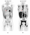
11-year-old female with stage IVA Hodgkin lymphoma (patient 1) demonstrating increased uptake in a right subcarinal lymph node. Biopsy revealed atypical lymphoid hyperplasia.
Figure 3
Figure 3
18-year-old female with stage II Hodgkin lymphoma (patient 4) demonstrating increased uptake in a left prevascular lymph node on FDG PET/CT (a, b and e, f) with 18F-FDG SUV 3.0 and no uptake on 18F-FLT/CT (c, d and g, h). Follow-up 18F-FDG PET/CT performed three (3) months later (not shown) again showed evidence of increased metabolic activity in the prevascular region in the upper mediastinum with SUV of 3.1 and not significantly changed compared to the previous study, and no other focus of increased 18F-FDG uptake to suggest disease progression.

References

    1. Bading J. R., Shields A. F. Imaging of cell proliferation: status and prospects. Journal of Nuclear Medicine. 2008;49(supplement 2):64S–80S. doi: 10.2967/jnumed.107.046391. - DOI - PubMed
    1. Shields A. F. Positron emission tomography measurement of tumor metabolism and growth: its expanding role in oncology. Molecular Imaging and Biology. 2006;8(3):141–150. doi: 10.1007/s11307-006-0039-2. - DOI - PubMed
    1. Shields A. F., Grierson J. R., Dohmen B. M., et al. Imaging proliferation in vivo with [F-18]FLT and positron emission tomography. Nature Medicine. 1998;4(11):1334–1336. doi: 10.1038/3337. - DOI - PubMed
    1. Choi S. J., Kim J. S., Kim J. H., et al. [18F]3′-deoxy-3′-fluorothymidine PET for the diagnosis and grading of brain tumors. European Journal of Nuclear Medicine and Molecular Imaging. 2005;32(6):653–659. doi: 10.1007/s00259-004-1742-3. - DOI - PubMed
    1. Gilles R., Vogel W. V., Gidding C. E. M., Janssens G. O. R. J., Van Der Vliet T. M., Oyen W. J. G. 18F-fluoro-L-thymidine-PET for the evaluation of primary brain tumours in children: a report of three cases. Nuclear Medicine Communications. 2010;31(6):482–487. doi: 10.1097/mnm.0b013e328318dc18. - DOI - PubMed

LinkOut - more resources