. 2016:2016:3067106.
doi: 10.1155/2016/3067106. Epub 2016 May 24.

Radiographic Follow-Up during Orthodontic Treatment for Early Diagnosis of Sequential Supernumerary Teeth

Affiliations

Radiographic Follow-Up during Orthodontic Treatment for Early Diagnosis of Sequential Supernumerary Teeth

Selma Sano Suga et al. Case Rep Dent. 2016.

Abstract

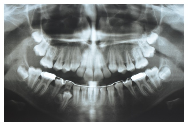
Most supernumerary teeth are impacted and asymptomatic. Objective. The aim of this paper is to describe two cases of sequential development of supernumerary teeth in the mandibular premolar region, identified during orthodontic treatment. Reports. The first case describes the radiographic follow-up of a female patient that presented a supernumerary tooth at the age of 9 years and 10 months in the right mandibular premolar region, followed by a further supernumerary tooth in the left mandibular premolar region identified at the age of 11 years and 3 months. In the second case, the radiographic follow-up of a male patient demonstrated 3 supernumerary teeth in the premolar region at the age of 16 years. During orthognathic surgery planning at the age of 20 years and 5 months, a supplemental supernumerary tooth was found in the left mandibular region. Conclusion. Considering the late developing of supernumerary premolars, appropriate follow-up with panoramic radiographs of patients with previous experience of supernumerary teeth is essential for early diagnosis of supplemental premolars to prevent possible complications.


Figures

Figure 1
Figure 1
(a) Panoramic radiograph of the patient aged 7 years and 3 months and (b) 9 years and 4 months with no evidence of supernumerary teeth.
Figure 2
Figure 2
Periapical radiograph of the patient aged 9 years and 10 months showing early formation of a supernumerary tooth in the right mandibular premolar region.
Figure 3
Figure 3
Panoramic radiograph of the patient aged 10 years 5 months showing early formation of a supernumerary tooth in the right mandibular premolar region.
Figure 4
Figure 4
Panoramic radiograph of the patient aged 11 years and 3 months showing the early formation of a supplemental supernumerary tooth in the left mandibular premolar region.
Figure 5
Figure 5
Panoramic radiographs of the patient aged (a) 12 years and 5 months and (b) 13 years and 7 months with no evidence of further supernumerary teeth.
Figure 6
Figure 6
Panoramic radiographs of the patient aged (a) 9 years and 11 months and (b) 10 years and 4 months with no evidence of supernumerary teeth.
Figure 7
Figure 7
Panoramic radiograph of the patient aged 11 years and 10 months with no evidence of supernumerary teeth.
Figure 8
Figure 8
Panoramic radiograph of the patient aged 16 years showing the early formation of 3 supernumerary teeth in the right and left mandibular premolar region and in the left maxillary premolar region.
Figure 9
Figure 9
Periapical radiograph of the patient showing a supernumerary tooth in the left maxillary region.
Figure 10
Figure 10
Panoramic radiograph of the patient aged 18 years after the extraction of all supernumerary teeth and third molars.
Figure 11
Figure 11
Panoramic radiograph of the patient aged 20 years and 5 months showing a supplemental supernumerary tooth in the left mandibular premolar region.

References

    1. Silva Filho O. G., Picolli V. D., Oliveira G. A. G., Bertoz F. A. Pré-molares supranumerários tardios: intercorrência remota no período pós-tratamento ortodôntico. Revista Clínica de Ortodontia Dental Press. 2010;8(6):52–59.
    1. Vahid-Dastjerdi E., Borzabadi-Farahani A., Mahdian M., Amini N. Supernumerary teeth amongst Iranian orthodontic patients. A retrospective radiographic and clinical survey. Acta Odontologica Scandinavica. 2011;69(2):125–128. doi: 10.3109/00016357.2010.539979. - DOI - PubMed
    1. Hyun H.-K., Lee S.-J., Ahn B.-D., et al. Nonsyndromic multiple mandibular supernumerary premolars. Journal of Oral and Maxillofacial Surgery. 2008;66(7):1366–1369. doi: 10.1016/j.joms.2007.08.028. - DOI - PubMed
    1. Anegundi R. T., Tavargeri A., Indushekar K. R., Sudha P. Sequential development of multiple supplemental premolars. Four-year follow-up report. The New York State Dental Journal. 2008;74(1):46–49. - PubMed
    1. Solares R., Romero M. I. Supernumerary premolars: a literature review. Pediatric Dentistry. 2004;26(5):450–458. - PubMed

LinkOut - more resources