

Review
doi: 10.3390/cells5020027.
Under Pressure: Mechanical Stress Management in the Nucleus
Affiliations
- PMID: 27314389
- PMCID: PMC4931676
- DOI: 10.3390/cells5020027
Item in Clipboard
Review
Under Pressure: Mechanical Stress Management in the Nucleus
Cells.
.
Abstract
Cells are constantly adjusting to the mechanical properties of their surroundings, operating a complex mechanochemical feedback, which hinges on mechanotransduction mechanisms. Whereas adhesion structures have been shown to play a central role in mechanotransduction, it now emerges that the nucleus may act as a mechanosensitive structure. Here, we review recent advances demonstrating that mechanical stress emanating from the cytoskeleton can activate pathways in the nucleus which eventually impact both its structure and the transcriptional machinery.
Keywords: LINC; lamin; lamina; mechanical stress; mechanotransduction; nucleoskeleton; nucleus.
Figures
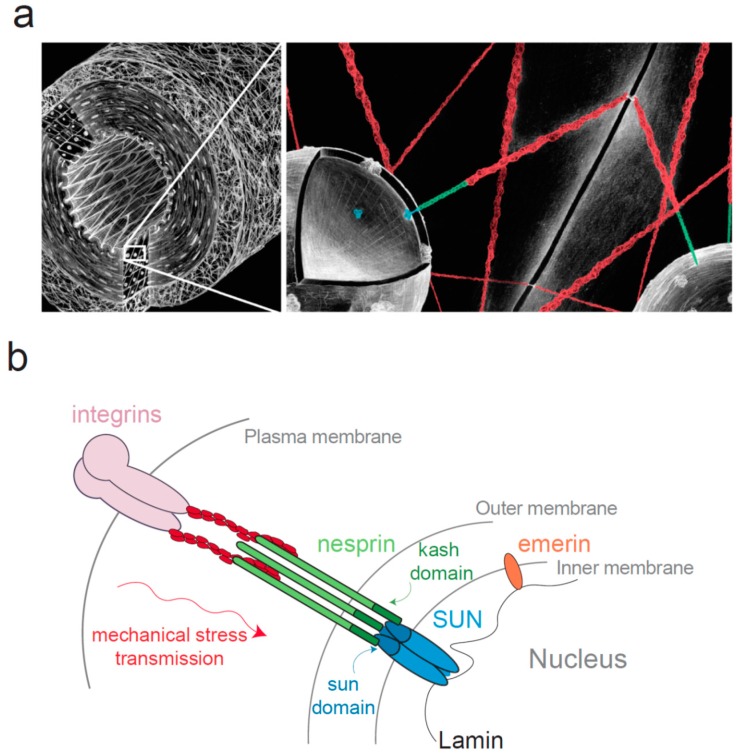
The Linker of Nucleoskeleton and Cytoskeleton (LINC) complex connects the nucleus to the cytoskeleton. (a) Diagram of a blood vessel (left panel) at two different scales, demonstrating the mechanical continuum that exists between the cell surface adhesion and the nucleus; (b) Schematic representation of the LINC complex. This complex consists in SUN proteins anchored in the inner nuclear membrane (INM) and nesprins anchored in the outer nuclear membrane (ONM). SUN domain was shown to organize in a trimeric fashion to bind three KASH peptides [50,51].
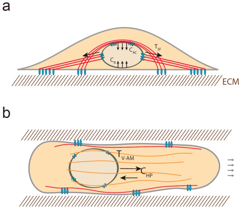
Mechanical stress experienced by the nucleus. (a) Diagram of a stationary cell. When cells are cultured on 2D surfaces, the nucleus can be subjected to tensional forces emanating from stress fibers (TSF) and compressive forces due to the actin cap (CAC) structures and the resistance of the surface (CR). The red solid lines represent the actin filaments; (b) In 3D, cells may also experience both tension, generated by vimentin-associated actomyosin filaments (orange structures) (TV-AM) and compression resulting from the high pressure of the anterior compartment (CHP).
References
-
- Thompson D. On Growth and Form. Cambridge University Press; New York, NY, USA: 1945.
-
- Austen K., Kluger C., Freikamp A., Chrostek-Grashoff A., Grashoff C. Generation and analysis of biosensors to measure mechanical forces within cells. Methods Mol. Biol. Clifton. 2013;1066:169–184. - PubMed
Publication types
Grants and funding
LinkOut - more resources
Full Text Sources
Other Literature Sources
