Development and Progression of Interstitial Lung Abnormalities in the Framingham Heart Study
- PMID: 27314401
- PMCID: PMC5215030
- DOI: 10.1164/rccm.201512-2523OC
Development and Progression of Interstitial Lung Abnormalities in the Framingham Heart Study
Abstract
Rationale: The relationship between the development and/or progression of interstitial lung abnormalities (ILA) and clinical outcomes has not been previously investigated.
Objectives: To determine the risk factors for, and the clinical consequences of, having ILA progression in participants from the Framingham Heart Study.
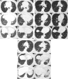
Methods: ILA were assessed in 1,867 participants who had serial chest computed tomography (CT) scans approximately 6 years apart. Mixed effect regression (and Cox) models were used to assess the association between ILA progression and pulmonary function decline (and mortality).
Measurements and main results: During the follow-up period 660 (35%) participants did not have ILA on either CT scan, 37 (2%) had stable to improving ILA, and 118 (6%) had ILA with progression (the remaining participants without ILA were noted to be indeterminate on at least one CT scan). Increasing age and increasing copies of the MUC5B promoter polymorphism were associated with ILA progression. After adjustment for covariates, ILA progression was associated with a greater FVC decline when compared with participants without ILA (20 ml; SE, ±6 ml; P = 0.0005) and with those with ILA without progression (25 ml; SE, ±11 ml; P = 0.03). Over a median follow-up time of approximately 4 years, after adjustment, ILA progression was associated with an increase in the risk of death (hazard ratio, 3.9; 95% confidence interval, 1.3-10.9; P = 0.01) when compared with those without ILA.
Conclusions: These findings demonstrate that ILA progression in the Framingham Heart Study is associated with an increased rate of pulmonary function decline and increased risk of death.
Keywords: MUC5B; idiopathic pulmonary fibrosis; interstitial lung abnormalities; interstitial lung disease; progression.
Figures


Comment in
-
Subclinical Interstitial Lung Abnormalities: Toward the Early Detection of Idiopathic Pulmonary Fibrosis?Am J Respir Crit Care Med. 2016 Dec 15;194(12):1445-1446. doi: 10.1164/rccm.201607-1363ED. Am J Respir Crit Care Med. 2016. PMID: 27976940 Free PMC article. No abstract available.
References
-
- Lederer DJ, Enright PL, Kawut SM, Hoffman EA, Hunninghake G, van Beek EJ, Austin JH, Jiang R, Lovasi GS, Barr RG. Cigarette smoking is associated with subclinical parenchymal lung disease: the Multi-Ethnic Study of Atherosclerosis (MESA)-lung study. Am J Respir Crit Care Med. 2009;180:407–414. - PMC - PubMed
-
- Tsushima K, Sone S, Yoshikawa S, Yokoyama T, Suzuki T, Kubo K. The radiological patterns of interstitial change at an early phase: over a 4-year follow-up. Respir Med. 2010;104:1712–1721. - PubMed
Publication types
MeSH terms
Grants and funding
- U01 HL105371/HL/NHLBI NIH HHS/United States
- T32 HL007633/HL/NHLBI NIH HHS/United States
- R21 HL120770/HL/NHLBI NIH HHS/United States
- R01 HL130974/HL/NHLBI NIH HHS/United States
- UH2 HL123442/HL/NHLBI NIH HHS/United States
- R01 HL116473/HL/NHLBI NIH HHS/United States
- R01 HL111024/HL/NHLBI NIH HHS/United States
- N01 HC025195/HL/NHLBI NIH HHS/United States
- I01 BX001534/BX/BLRD VA/United States
- R01 HL107246/HL/NHLBI NIH HHS/United States
- R01 HL097163/HL/NHLBI NIH HHS/United States
- K25 HL104085/HL/NHLBI NIH HHS/United States
- P01 HL114501/HL/NHLBI NIH HHS/United States
- R01 HL122464/HL/NHLBI NIH HHS/United States
- P01 HL092870/HL/NHLBI NIH HHS/United States
- R33 HL120770/HL/NHLBI NIH HHS/United States
- UH3 HL123442/HL/NHLBI NIH HHS/United States
- R01 HL129920/HL/NHLBI NIH HHS/United States
- K23 CA157631/CA/NCI NIH HHS/United States
LinkOut - more resources
Full Text Sources
Other Literature Sources
Medical
Research Materials

