. 2016 Oct;10(4):178-86.
doi: 10.1080/19336934.2016.1195938. Epub 2016 Jun 17.

Diaphanous regulates SCAR complex localization during Drosophila myoblast fusion

Affiliations

Diaphanous regulates SCAR complex localization during Drosophila myoblast fusion

Su Deng et al. Fly (Austin). 2016 Oct.

Abstract

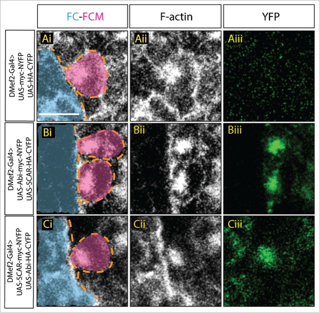
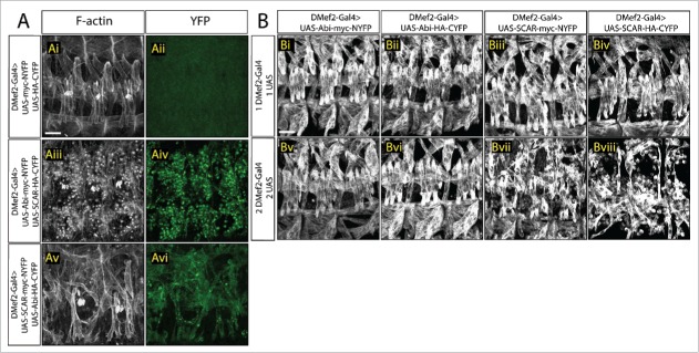
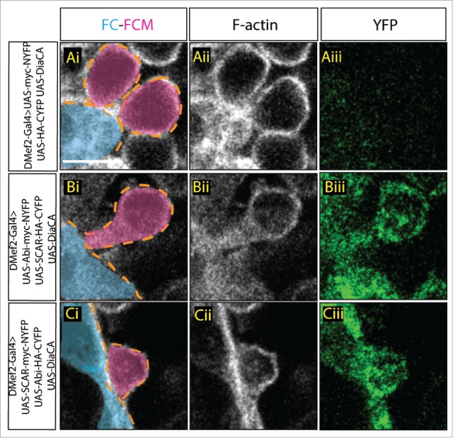
From Drosophila to man, multinucleated muscle cells form through cell-cell fusion. Using Drosophila as a model system, researchers first identified, and then demonstrated, the importance of actin cytoskeletal rearrangements at the site of fusion. These actin rearrangements at the fusion site are regulated by SCAR and WASp mediated Arp2/3 activation, which nucleates branched actin networks. Loss of SCAR, WASp or both leads to defects in myoblast fusion. Recently, we have found that the actin regulator Diaphanous (Dia) also plays a role both in organizing actin and in regulating Arp2/3 activity at the fusion site. In this Extra View article, we provide additional data showing that the Abi-SCAR complex accumulates at the fusion site and that excessive SCAR activity impairs myoblast fusion. Using constitutively active Dia constructs, we provide additional evidence that Dia functions upstream of SCAR activity to regulate actin dynamics at the fusion site and to localize the Abi-SCAR complex.

Keywords: Abi; Arp2/3; Diaphanous; SCAR/WAVE; actin; myoblast fusion.


Figures

Figure 1.
Figure 1.
Visualization of Abi-SCAR complex formation using split YFP during myoblast fusion. Stage 15 embryo stained for F-actin (phalloidin, white) to label fusion site, and YFP (GFP antibody, green) to detect YFP reconstitution, FCM (magenta, false colored), and FC/myotube (turquoise, false colored). (Ai-Aiii) To visualize the background fluorescent level, UAS-myc-NYFP and UAS-HA-CYFP were expressed in the muscles under the control of muscles specific driver DMef2-Gal4. (Bi-Biii) UAS-Abi-myc-NYFP and UAS-SCAR-HA-CYFP; or (Ci-Ciii) UAS-SCAR-myc-NYFP and UAS-Abi-HA-CYFP were expressed in the muscles under the control of DMef2-Gal4. The reconstituted YFP signals indicate sites of Abi-SCAR interaction. Scale bar: 5 μm.
Figure 2.
Figure 2.
Increased SCAR activity results in fusion block. A. Three hemisegments from a stage 16 embryo. Embryos are stained for F-actin (phalloidin, white) to show the muscle pattern, and YFP (GFP antibody, green) to detect YFP reconstitution. (Ai–Aii) In control embryos, UAS-myc-NYFP and UAS-HA-CYFP were expressed in muscles under the control of DMef2-Gal4. Phalloidin staining shows wild-type muscle pattern. Background fluorescent level was visualized with antibody against GFP. UAS-Abi-myc-NYFP and UAS-SCAR-HA-CYFP (Aiii–Aiv) or UAS-SCAR-myc-NYFP and UAS-Abi-HA-CYFP (Av–Avi) were expressed in muscles under the control of DMef2-Gal4. Phalloidin staining shows impaired fusion and the actin focus at the fusion site. YFP shows the localization of Abi-SCAR interaction. (B) Muscle pattern from stage 16 embryos (antibody against Myosin Heavy Chain. white). Three hemisegments are shown from each embryo. One (Bi–Biv) or 2 copies (Bv–Bviii) of split-YFP labeled Abi or SCAR were expressed in the muscles under the control of DMef2-Gal4. Abi overexpression does not change muscle pattern. Increased expression of SCAR results in myoblast fusion block in a dosage dependent manner. Scale bar: 20 μm.
Figure 3.
Figure 3.
Constitutively active Dia changes Abi-SCAR localization and actin structure at the fusion site. Stage 16 embryo stained for F-actin (phalloidin, white) and YFP (GFP antibody, green), FCM (magenta, false colored), FC/myotube (turquoise/false colored). (Ai–Aiii) As control, constitutively active Dia (UAS-Dia.CA) was expressed together with UAS-myc-NYFP and UAS-HA-CYFP in the muscles under the control of DMef2-Gal4. Fusion is blocked in this context. Background YFP fluorescent level was visualized with antibody against GFP. (B-C) UAS-Dia.CA was expressed together with UAS-Abi-myc-NYFP and UAS-SCAR-HA-CYFP (Bi-Biii) or UAS-SCAR-myc-NYFP and UAS-Abi-HA-CYFP (Ci-Ciii) in the muscles. Actin morphology at the fusion site was visualized by Phalloidin staining of F-actin. Abi-SCAR complex was visualized by YFP reconstitution and was labeled by antibody against GFP. Scale bar: 5 μm.

Comment on

  • doi: 10.1371/journal.pgen.1005381

References

    1. Kim JH, Jin P, Duan R, Chen EH. Mechanisms of myoblast fusion during muscle development. Curr Opin Genet Dev 2015; 32:162-70; PMID:25989064; http://dx.doi.org/10.1016/j.gde.2015.03.006 - DOI - PMC - PubMed
    1. Abmayr SM, Pavlath GK. Myoblast fusion: lessons from flies and mice. Development 2012; 139:641-56; PMID:22274696; http://dx.doi.org/10.1242/dev.068353 - DOI - PMC - PubMed
    1. Simionescu A, Pavlath GK. Molecular mechanisms of myoblast fusion across species. Adv Exp Med Biol 2011; 713:113-35; PMID:21432017; http://dx.doi.org/10.1007/978-94-007-0763-4_8 - DOI - PubMed
    1. Rai M, Nongthomba U, Grounds MD. Skeletal muscle degeneration and regeneration in mice and flies. Curr Top Dev Biol 2014; 108:247-81; PMID:24512712; http://dx.doi.org/10.1016/B978-0-12-391498-9.00007-3 - DOI - PubMed
    1. Onel S-F, Rust MB, Jacob R, Renkawitz-Pohl R. Tethering membrane fusion: common and different players in myoblasts and at the synapse. J Neurogenet 2014; 28:302-15; PMID:24957080; http://dx.doi.org/10.3109/01677063.2014.936014 - DOI - PMC - PubMed

Publication types

MeSH terms

LinkOut - more resources