. 2016 Aug;14(2):1610-6.
doi: 10.3892/mmr.2016.5405. Epub 2016 Jun 16.

Jia-Shen decoction-medicated serum inhibits angiotensin-II induced cardiac fibroblast proliferation via the TGF-β1/Smad signaling pathway

Affiliations

Jia-Shen decoction-medicated serum inhibits angiotensin-II induced cardiac fibroblast proliferation via the TGF-β1/Smad signaling pathway

Lin Cui et al. Mol Med Rep. 2016 Aug.

Abstract

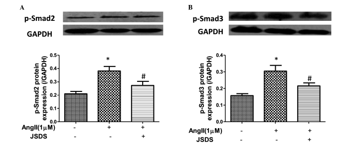
Jia-Shen decoction (JSD) is a traditional Chinese medicine, which is used widely to treat chronic heart failure. However, the underlying mechanism remains to be elucidated. The present study aimed to investigate the mechanism underlying the effects of JSD on cardiac fibroblast (CF) proliferation and differentiation. The CFs were obtained from the hearts of neonatal (1‑3‑day old) Sprague‑Dawley rats and treated with JSD-medicated serum (JSDS) with or without angiotensin II (Ang II). Cell proliferation was assessed using Cell Counting Kit‑8 reagent. In addition, the mRNA expression levels of transforming growth factor‑β1 (TGF‑β1) and phosphorylated small mothers against decapentaplegic (p‑Smad)2/3 and their protein expression levels were analyzed. CF proliferation was significantly increased in the Ang II‑treated group, compared with the control group (P<0.05). The expression levels of collagen, α‑smooth muscle actin, TGF‑β1 and p‑Smad2/3 were also increased in the Ang II‑treated group (P<0.05). Following JSDS treatment, the increased levels of collagen and cell proliferation were inhibited, and the increased expression levels of p‑Smad2 and p‑Smad3 were also inhibited (P<0.05). These data suggested that JSDS may inhibit CF proliferation via attenuating the TGF‑β1/Smad signaling pathway.


Figures

Figure 1
Figure 1
Characterization of primary cultured neonatal rat CFs. (A) Cell morphology of the first passage of neonatal rat CFs under an optical microscope. Original magnification, ×20. (B) Primary cultured CFs were subjected to immunofluorescence staining for vimentin to determine their purity. Red shows vimentin staining; DAPI is shown in blue (nuclear). Original magnification, ×40. CFs, cardiac fibroblasts.
Figure 2
Figure 2
Effects of different concentrations of Ang II on cell proliferation in CFs. (A) Neonatal rat CFs were treated with different concentrations of Ang II for 24 h. (B) CFs were treated with different concentration of Ang II for 48 h. Data are presented as the mean ± standard error of the mean. *P<0.05, compared with the non-Ang II-treated group. CFs, cardiac fibroblasts; Ang II, angiotensin II; OD, optical density.
Figure 3
Figure 3
JSDS inhibits Ang II-induced cell proliferation and reduces collagen content in CFs. (A) Neonatal rat CFs were treated with different concentrations of JSDS and Ang II (1 µM) for 24 h. The number of cells are presented as an OD value, determined using a cell count assay. (B) Content of hydroxyproline in cell culture medium. *P<0.05, compared with the control group; #P<0.05, compared with the Ang II-treated group. Data are presented as the mean ± standard error of the mean. CFs, cardiac fibroblasts; JSDS, Jia-Shen decoction-medicated serum; Ang II, angiotensin II; OD, optical density.
Figure 4
Figure 4
Effects of JSDS on Ang II-induced expression of collagen I in CFs. (A) mRNA expression levels of collagen I were assessed using reverse transcription-quantitative polymerase chain reaction analysis. (B) Fluorescence intensity of the expression of collagen I. (C) Cellular collagen I was visualized using fluorescein isothiocyanate-conjugated IgG in immunofluorescence staining. Representative images of the (a) control group, (b) Ang II-treated group. And (c) JSDS-treated group. Values are expressed as the mean ± standard error of the mean (n=3). *P<0.05, compared with the control group; #P<0.05, compared with the Ang II-treated group. Original magnification, ×20, with representative images shown; n=3. CFs, cardiac fibroblasts; JSDS, Jia-Shen decoction-medicated serum; Ang II, angiotensin II.
Figure 5
Figure 5
Effects of JSDS on Ang II-induced expression of α-SMA in CFs. (A) mRNA expression levels of α-SMA in the different groups were assessed using reverse trancsription-quantitative polymerase chain reaction analysis. (B) Fluorescence intensity of the expression of α-SMA. (C) Cellular α-SMA was visualized using fluorescein isothiocyanateconjugated IgG by immunofluorescence staining. Representative images of the (a) control group, the (b) AngII-treated group and the (c) JSDS-treated group. Values are expressed as the mean ± standard error of the mean (n=3). *P<0.05, compared with the control group; #P<0.05, compared with the Ang II-treated group. Original magnification, ×20, with representative images shown; n=3. CFs, cardiac fibroblasts; JSDS, Jia-Shen decoction-medicated serum; α-SMA, α-smooth muscle actin; Ang II, angiotensin II.
Figure 6
Figure 6
JSDS inhibits Ang II-induced relative mRNA and protein levels of TGF-β1 in CFs. (A) mRNA expression levels of TGF-β1 were assessed using reverse transcription-quantitative polymerase chain reaction analysis. Data were normalized to GAPDH mRNA. (B) Cell lysates were analyzed to determine the protein expression levels of TGF-β1 using western blot analysis. Values are expressed as the mean ± standard error of the mean (n=3). *P<0.05, compared with the control group; #P<0.05, compared with the Ang II-treated group. CFs, cardiac fibroblasts; JSDS, Jia-Shen decoction-medicated serum; TGF-β1, transforming growth factor-β1; Ang II, angiotensin II.
Figure 7
Figure 7
JSDS inhibits Ang II-induced expression of p-Smad2 and p-Smad3 in CFs. (A) Protein expression levels of p-Smad2 were determined using western blot analysis. (B) Protein expression levels of p-Smad3 were determined using western blot analysis. Values are expressed as the mean ± standard error of the mean (n=3). *P<0.05, compared with the control group; #P<0.05, compared with the Ang II-treated group. CFs, cardiac fibroblasts; JSDS, Jia-Shen decoction-medicated serum; p-Smad, phosphorylated small mothers against decapentaplegic; Ang II, angiotensin II.

References

    1. Wynn TA, Ramalingam TR. Mechanisms of fibrosis: Therapeutic translation for fibrotic disease. Nat Med. 2012;18:1028–1040. doi: 10.1038/nm.2807. - DOI - PMC - PubMed
    1. Crabos M, Roth M, Hahn AW, Erne P. Characterization of angiotensin II receptors in cultured adult rat cardiac fibroblasts. Coupling to signaling systems and gene expression. J Clin Invest. 1994;93:2372–2378. doi: 10.1172/JCI117243. - DOI - PMC - PubMed
    1. Dostal DE, Booz GW, Baker KM. Angiotensin II signalling pathways in cardiac fibroblasts: Conventional versus novel mechanisms in mediating cardiac growth and function. Mol Cell Biochem. 1996;157:15–21. doi: 10.1007/BF00227876. - DOI - PubMed
    1. Sadoshima J, Izumo S. Molecular characterization of angiotensin II-induced hypertrophy of cardiac myocytes and hyperplasia of cardiac fibroblasts. Critical role of the AT1 receptor subtype. Circ Res. 1993;73:413–423. doi: 10.1161/01.RES.73.3.413. - DOI - PubMed
    1. Zhou G, Kandala JC, Tyagi SC, Katwa LC, Weber KT. Effects of angiotensin II and aldosterone on collagen gene expression and protein turnover in cardiac fibroblasts. Mol Cell Biochem. 1996;154:171–178. doi: 10.1007/BF00226785. - DOI - PubMed

MeSH terms