. 2016 Jun 16;165(7):1762-1775.
doi: 10.1016/j.cell.2016.06.001.

Microbial Reconstitution Reverses Maternal Diet-Induced Social and Synaptic Deficits in Offspring

Affiliations

Microbial Reconstitution Reverses Maternal Diet-Induced Social and Synaptic Deficits in Offspring

Shelly A Buffington et al. Cell. .

Abstract

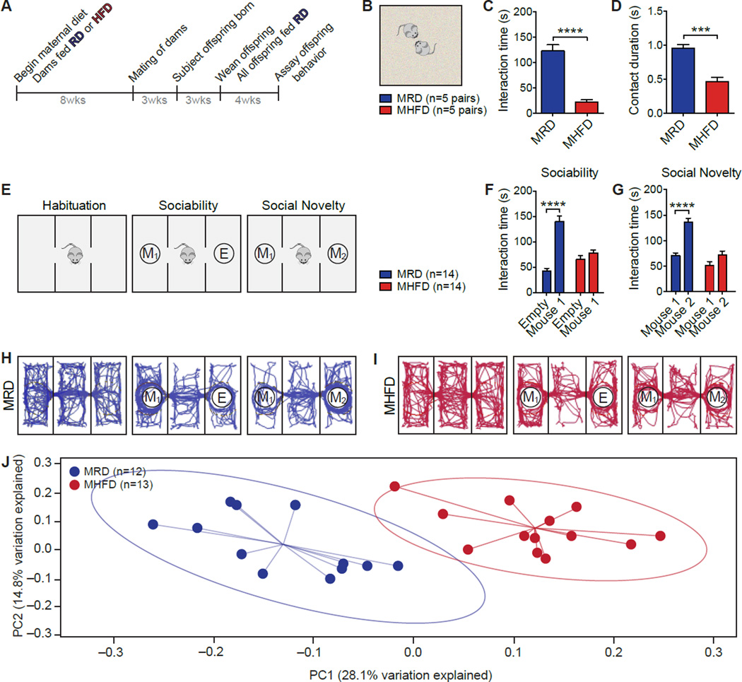
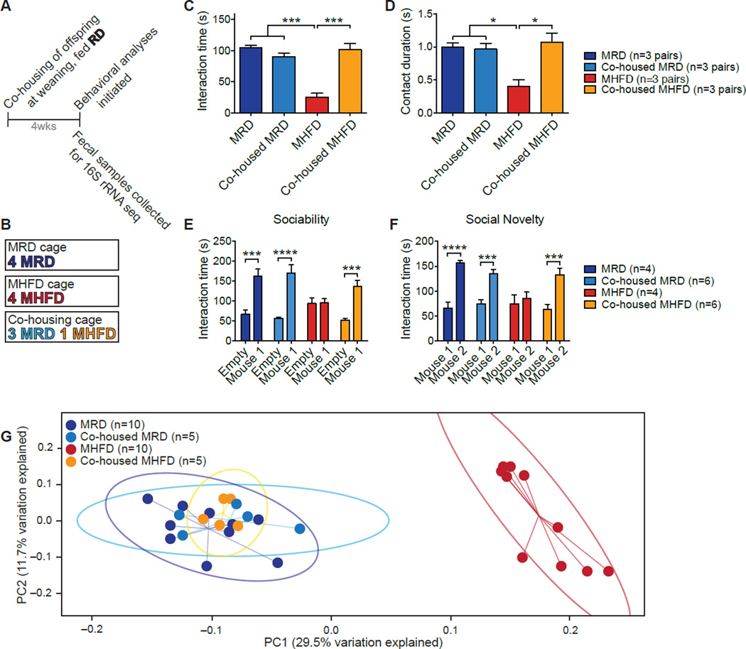
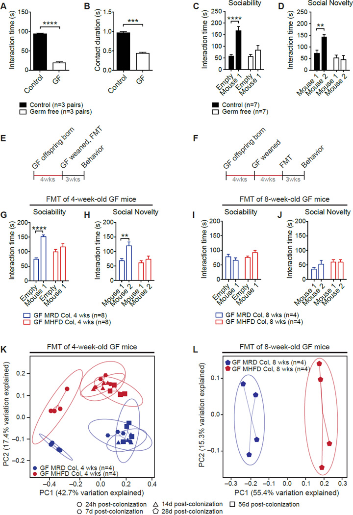
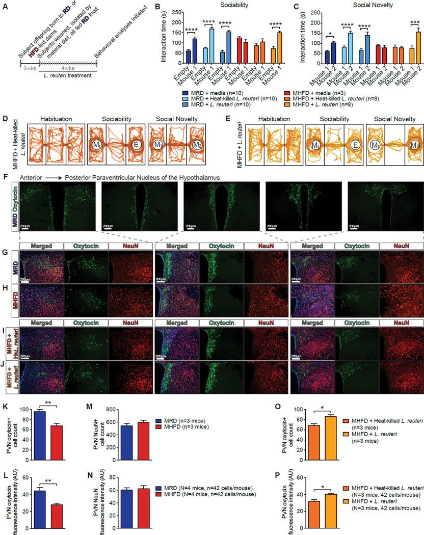
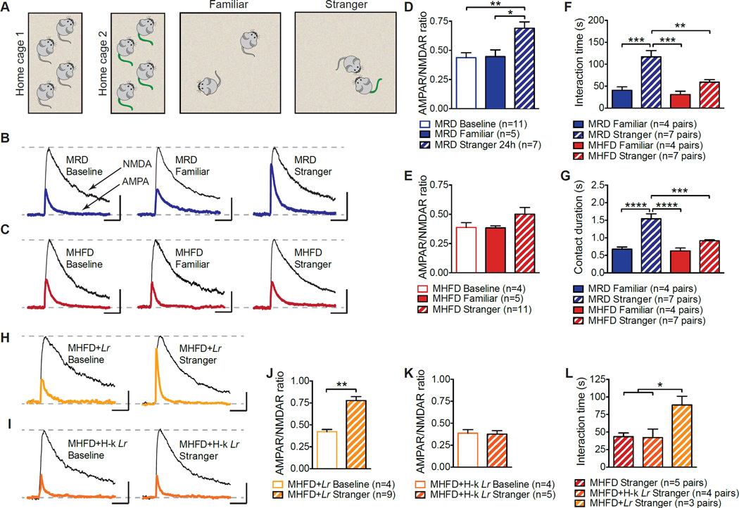
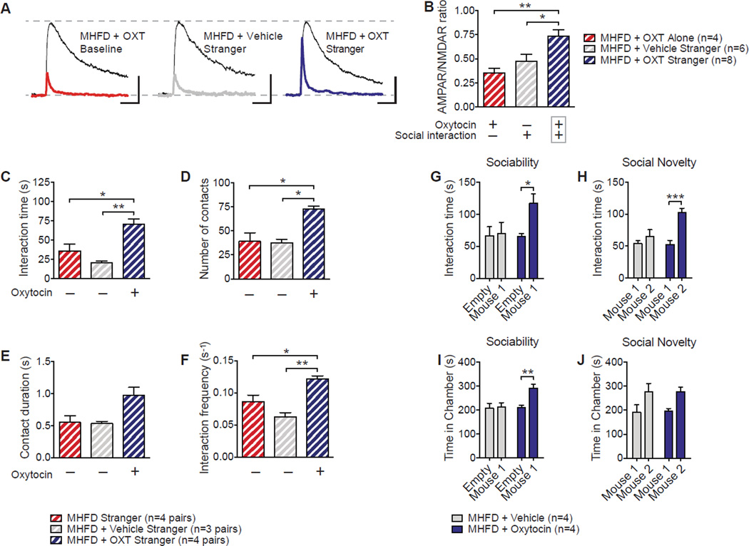
Maternal obesity during pregnancy has been associated with increased risk of neurodevelopmental disorders, including autism spectrum disorder (ASD), in offspring. Here, we report that maternal high-fat diet (MHFD) induces a shift in microbial ecology that negatively impacts offspring social behavior. Social deficits and gut microbiota dysbiosis in MHFD offspring are prevented by co-housing with offspring of mothers on a regular diet (MRD) and transferable to germ-free mice. In addition, social interaction induces synaptic potentiation (LTP) in the ventral tegmental area (VTA) of MRD, but not MHFD offspring. Moreover, MHFD offspring had fewer oxytocin immunoreactive neurons in the hypothalamus. Using metagenomics and precision microbiota reconstitution, we identified a single commensal strain that corrects oxytocin levels, LTP, and social deficits in MHFD offspring. Our findings causally link maternal diet, gut microbial imbalance, VTA plasticity, and behavior and suggest that probiotic treatment may relieve specific behavioral abnormalities associated with neurodevelopmental disorders. VIDEO ABSTRACT.

Keywords: autism; dysbiosis; high-fat diet (HFD); long-term potentiation (LTP); neurodevelopmental disorders; probiotic; ventral tegmental area (VTA).


Figures

Figure 1
Figure 1. Social Deficits and Dysbiosis of the Gut Microbiota in MHFD Offspring
A, Schematic of the maternal diet regimen and breeding. B, Schematic of the reciprocal social interaction task. C–D, MHFD offspring showed reduced reciprocal interaction (C, P<0.0001, t=7.90; D, P<0.001, t=5.89). E, Schematic of the three-chamber social interaction task. F–G, In the sociability test, MRD offspring spent more time interacting with a mouse than with an empty wire cage (F, P<0.0001, t=8.817), whereas MHFD offspring showed no preference for the mouse (F, P=0.48, t=1.19; Maternal diet effect F1,52=6.08, P<0.05). In the social novelty test, unlike MRD (G, P<0.0001, t=6.68), MHFD offspring had no preference for interacting with a novel versus a familiar mouse (G, P=0.086, t=2.08; Maternal diet effect F1,52=34.96, P<0.0001). H–I, Representative exploratory activity of MRD (H) and MHFD (I) offspring in the three-chamber test. J, Principal coordinates analysis (PCoA) of unweighted UniFrac distances from the averaged rarefied 16S rRNA gene dataset (n=1,000 rarefactions; 7,617 reads/sample) showed that MRD samples clustered separately from MHFD samples (P<0.001, R2=0.37). Plots show mean ± SEM. See also Figures S1–S2.
Figure 2
Figure 2. Co-housing MHFD with MRD Offspring Rescues both Social Dysfunction and the Microbiota Phylogenetic Profile of MHFD Mice
A, Schematic of the co-housing experiment. B, MRD and MHFD offspring were weaned into one of three cage compositions. C–G, Social interaction time (C, MRD vs. MHFD P<0.001, t=9.30; MRD vs. co-housed MHFD P>0.99, t=0.31; MHFD vs. co-housed MHFD P<0.001, t=7.99; P<0.0001, F3,8=30.51) and contact duration (D, MRD vs. MHFD P<0.05, t=4.13; MRD vs. co-housed MHFD P>0.99, t=0.46; MHFD vs. co-housed MHFD P<0.05, t=4.59; P<0.001, F3,8=9.01) in the reciprocal interaction test; social interaction times in the sociability (E, MRD P<0.001, t=4.36; MHFD P>0.99, t=0.078; Co-housed MRD P<0.0001, t=6.33; Co-housed MHFD P<0.001, t=4.78; Maternal diet/Housing/Interaction effect F3,32=6.13, P<0.01) and social novelty tests (F, MRD P<0.0001, t=5.12; MHFD P>0.99, t=0.60; Co-housed MRD P<0.001, t=4.20; Co-housed MHFD P<0.001, t=4.76; Maternal diet/Housing/Interaction effect F3,32=4.37, P<0.01), as well as UniFrac-based phylogenetic clustering (G, P<0.001, R2=0.552; n=1,000 rarefactions; 3,390 reads/sample), are all restored in MHFD offspring co-housed with MRD mice. Plots show mean ± SEM. See also Figure S3.
Figure 3
Figure 3. Fecal Microbiota from MRD, but not MHFD, Offspring Improves Germ-Free (GF) Recipient Social Behavior
A–D, GF mice show reduced reciprocal social interaction (A, P<0.0001, t=22.73; B, P<0.001, t=11.31) and deficits in sociability (C, Control P<0.0001, t=5.30, GF P>0.99, t=0.39; Main group effect F1,24=21.98, P<0.0001) and preference for social novelty (D, Control P<0.01, t=3.64, GF P=0.39, t=1.33; Main group effect F1,24=5.29, P<0.05). Schematic of fecal microbiota transplant (FMT) at four (E) and eight weeks of age (F). G–H, FMT from MRD, but not MHFD, offspring at weaning restored both GF sociability (G, GFMRDCol P<0.0001, t=6.66; GFMHFDCol P=0.35, t=1.40; Donor effect F1,28=32.44, P<0.0001) and preference for social novelty (H, GFMRDCol P<0.01, t=3.60; GFMHFDCol P=0.81, t=0.84; Donor effect F1,28=9.86, P<0.01). I–J, At eight weeks, FMT from either MRD or MHFD donors failed to improve sociability (I, GFMRDCol P=0.51, t=1.20; GFMHFDCol P=0.28, t=1.58; Donor effect F1,12=0.07, P=0.79) or preference for social novelty in GF mice (J, GFMRDCol P=0.48, t=1.23; GFMHFDCol P>0.99, t=0.043; Donor effect F1,12=0.71, P=0.42). K–L, PCoA of unweighted UniFrac distances based on the 16S rRNA gene sequencing dataset from GF recipients of stools from either MRD or MHFD donors at four (K, P=0.001, R2=0.83; n=1,000 rarefactions; 4,628 reads/sample) or eight (L, P<0.001, R2=0.77; n=1,000 rarefactions; 4,805 reads/sample) weeks of age. Plots show mean ± SEM. See also Figure S4.
Figure 4
Figure 4. Selective Treatment with Lactobacillus (L.) reuteri Restores Social Deficits and Oxytocin Levels in MHFD Offspring
A, Schematic of L. reuteri–treatment. B,C, Unlike resuspension media (B, P>0.99, t=1.03; c, P>0.99, t=0.40) or heat-killed L. reuteri (B, P>0.99, t=1.35; c, P>0.99, t=0.21), administration of live L. reuteri in the drinking water rescued sociability (B, P<0.0001, t=5.98) and preference for social novelty (C, P<0.001, t=5.01) in MHFD offspring (B, Treatment effect F1,86=87.53, P<0.0001; C, Treatment effect F1,86=30.24, P<0.0001). D–E, Representative track plots showing exploratory activity. F, Representative images of control oxytocin immunoreactivity at different anteroposterior levels of the PVN. G–J, Oxytocin immunoreactivity in the PVN of MRD (G), MHFD (H), heat-killed L. reuteri-treated MHFD (I), and live L. reuteri-treated MHFD offspring (J). K–N, Oxytocin immunoreactive cell number (K, P<0.01, t=4.76) and oxytocin immunofluorescence intensity (L, P<0.01, t=3.80) were reduced in the PVN of MHFD versus MHFD mice. In the PVN of MRD and MHFD offspring, NeuN cell number immunoreactivity (M, P=0.34, t=1.09) and immunofluorescence intensity (N, P=0.79, t=0.28) were similar. O–P, Relative to treatment with heat-killed L. reuteri, treatment with live L. reuteri significantly increased oxytocin-positive cell number (O, P<0.05, t=2.93) and oxytocin immunofluorescence intensity (P, P<0.05, t=3.09) in the PVN of MHFD offspring. AU: arbitrary units. Plots show mean ± SEM. See also Figures S5–S6.
Figure 5
Figure 5. Reciprocal Social Interaction and Social Interaction-Induced LTP in MHFD Offspring VTA DA Neurons are Restored by L. reuteri
A, Schematic of the experimental design. B–E, LTP was measure 24 hours following reciprocal interaction with either a stranger or a familiar mouse. Only interaction with a stranger mouse induced LTP in MRD VTA DA neurons, as determined by increased AMPAR/NMDAR ratios (B,D, Baseline vs. Familiar P>0.99, t=0.12, Baseline vs. Stranger P<0.01, t=3.79; Familiar vs. Stranger P<0.05, t=3.03; F=8.03, P<0.01). In MHFD offspring, neither stranger nor familiar reciprocal interaction evoked LTP in VTA DA neurons (C,E, Baseline vs. Familiar P>0.99, t=0.035, Baseline vs. Stranger P=0.64, t=1.30; Familiar vs. Stranger P=0.50, t=1.45; F2,15=1.47, P=0.26). F–G, Whereas MRD mice spent more time interacting with a stranger than a familiar mouse (Familiar vs. Stranger; F, P<0.001, t=4.88; G, P<0.0001, t=5.87), MHFD mice did not (Familiar vs. Stranger; F, P=0.47, t=1.87; G, P=0.40, t=1.96) (F, F3,19=13.8, P<0.0001; G, F3,19=18.54, P<0.0001). H–K, Live (H,J, P<0.01, t=4.95), but not heat-killed L. reuteri (I,K, P=0.84, t=0.20), restored stranger interaction-evoked LTP in the VTA of MHFD offspring. L, Unlike heat-killed L. reuteri (MHFD vs. MHFD+Hk−Lr P>0.99, t=0.099), live L. reuteri restored reciprocal social interaction (MHFD vs. MHFD+Lr P<0.05, t=3.24; F2,9=6.45, P<0.05). Plots show mean ± SEM. See also Figure S7.
Figure 6
Figure 6. Oxytocin Restores Social Interaction-Induced VTA Plasticity and Social Behavioral Deficits in MHFD Offspring
A–B, LTP was measure 1–3 hours following a reciprocal interaction. Intranasal oxytocin administration rescued LTP in the VTA of MHFD offspring (B, MHFD+OXT Alone vs. MHFD+OXT Stranger P<0.01, t=3.66; MHFD+Vehicle Stranger vs. MHFD+OXT Stranger P<0.05, t=2.86; F2,15=7.97, P<0.01). C–F, Oxytocin restored reciprocal social interaction (C, MHFD vs. MHFD+Vehicle P=0.55, t=1.46; MHFD vs. MHFD+OXT P<0.05, t=3.62; MHFD+Vehicle vs. MHFD+OXT P<0.01, t=4.81; F2,8=12.82, P<0.01; D, MHFD vs. MHFD+Vehicle P>0.99, t=0.16; MHFD vs. MHFD+OXT P<0.05, t=4.075; MHFD+Vehicle vs. MHFD+OXT P<0.05, t=3.94; F2,8=10.97, P<0.01; E, MHFD vs. MHFD+Vehicle, P>0.99, t=0.11; MHFD vs. MHFD+OXT P=0.052, t=2.99; Treatment effect F2,8=5.87, P<0.05; F, MHFD vs. MHFD+Vehicle P=0.2, t=2.11; MHFD vs. MHFD+OXT P<0.05, t=3.43; Treatment effect F2,8=14.58, P<0.01), sociability (G, MHFD+Vehicle P>0.99, t=0.44; MHFD+Oxytocin P=0.24, t=1.74; Treatment effect F1,8=2.37, P=0.16; H, MHFD+Vehicle P>0.99, t=0.29; MHFD+Oxytocin P<0.01, t=3.50; Treatment effect F1,12=5.16, P<0.05) and preference for social novelty in MHFD offspring (I, MHFD+Vehicle P=0.65, t=1.05; MHFD+Oxytocin P<0.05, t=3.54; Treatment effect F1,8=10.54, P<0.05; J, MHFD+Vehicle P=0.50, t=1.25; MHFD+Oxytocin P=0.096, t=2.34; Treatment effect F(1,8)=6.41, P<0.05). Plots show mean ± SEM.

Comment in

References

    1. Aye IL, Rosario FJ, Powell TL, Jansson T. Adiponectin supplementation in pregnant mice prevents the adverse effects of maternal obesity on placental function and fetal growth. Proceedings of the National Academy of Sciences of the United States of America. 2015;112:12858–12863. - PMC - PubMed
    1. Benjamini Y, Drai D, Elmer G, Kafkafi N, Golani I. Controlling the false discovery rate in behavior genetics research. Behav Brain Res. 2001;125:279–284. - PubMed
    1. Bilder DA, Bakian AV, Viskochil J, Clark EA, Botts EL, Smith KR, Pimentel R, McMahon WM, Coon H. Maternal prenatal weight gain and autism spectrum disorders. Pediatrics. 2013;132:e1276–e1283. - PMC - PubMed
    1. Bolton JL, Bilbo SD. Developmental programming of brain and behavior by perinatal diet: focus on inflammatory mechanisms. Dialogues in clinical neuroscience. 2014;16:307–320. - PMC - PubMed
    1. Bravo JA, Forsythe P, Chew MV, Escaravage E, Savignac HM, Dinan TG, Bienenstock J, Cryan JF. Ingestion of Lactobacillus strain regulates emotional behavior and central GABA receptor expression in a mouse via the vagus nerve. Proceedings of the National Academy of Sciences of the United States of America. 2011;108:16050–16055. - PMC - PubMed

Publication types