. 2016 Dec;6(1):52.
doi: 10.1186/s13550-016-0207-6. Epub 2016 Jun 17.

A dual tracer (68)Ga-DOTANOC PET/CT and (18)F-FDG PET/CT pilot study for detection of cardiac sarcoidosis

Affiliations

A dual tracer (68)Ga-DOTANOC PET/CT and (18)F-FDG PET/CT pilot study for detection of cardiac sarcoidosis

Lars C Gormsen et al. EJNMMI Res. 2016 Dec.

Abstract

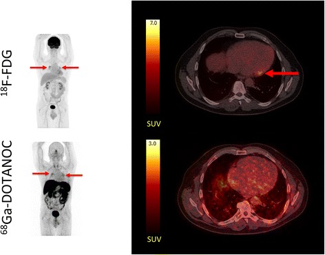
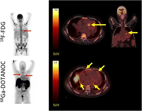
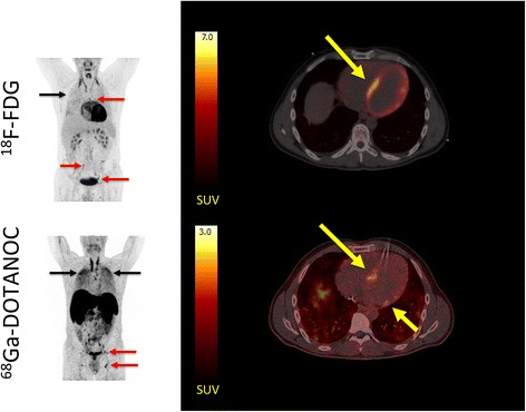
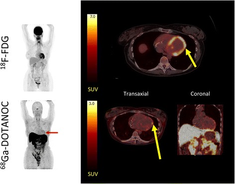
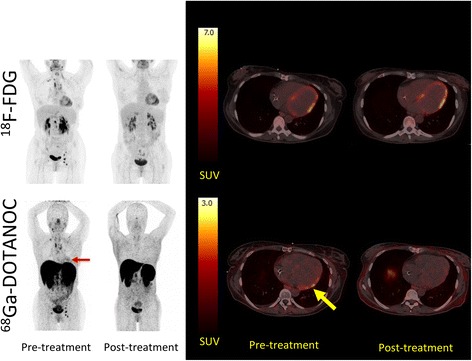
Background: Cardiac sarcoidosis (CS) is a potentially fatal condition lacking a single test with acceptable diagnostic accuracy. (18)F-FDG PET/CT has emerged as a promising imaging modality, but is challenged by physiological myocardial glucose uptake. An alternative tracer, (68)Ga-DOTANOC, binds to somatostatin receptors on inflammatory cells in sarcoid granulomas. We therefore aimed to conduct a proof-of-concept study using (68)Ga-DOTANOC to diagnose CS. In addition, we compared diagnostic accuracy and inter-observer variability of (68)Ga-DOTANOC vs. (18)F-FDG PET/CT.

Methods: Nineteen patients (seven female) with suspected CS were prospectively recruited and dual tracer scanned within 7 days. PET images were reviewed by four expert readers for signs of CS and compared to the reference standard (Japanese ministry of Health and Welfare CS criteria).

Results: CS was diagnosed in 3/19 patients. By consensus, 11/19 (18)F-FDG scans and 0/19 (68)Ga-DOTANOC scans were rated as inconclusive. The sensitivity of (18)F-FDG PET for diagnosing CS was 33 %, specificity was 88 %, PPV was 33 %, NPV was 88 %, and diagnostic accuracy was 79 %. For (68)Ga-DOTANOC, accuracy was 100 %. Inter-observer agreement was poor for (18)F-FDG PET (Fleiss' combined kappa 0.27, NS) and significantly better for (68)Ga-DOTANOC (Fleiss' combined kappa 0.46, p = 0.001).

Conclusions: Despite prolonged pre-scan fasting, a large proportion of (18)F-FDG PET/CT images were rated as inconclusive, resulting in low agreement among reviewers and correspondingly poor diagnostic accuracy. By contrast, (68)Ga-DOTANOC PET/CT had excellent diagnostic accuracy with the caveat that inter-observer variability was still significant. Nevertheless, (68)Ga-DOTANOC PET/CT looks very promising as an alternative CS PET tracer.

Trial registration: Current Controlled Trials NCT01729169 .

Keywords: Cardiac sarcoidosis; Heart failure; Inflammation; PET/CT; Somatostatin receptor imaging.


Figures

Fig. 1
Fig. 1
Typical 18F-FDG PET/CT false positive for cardiac sarcoidosis. Left panel: maximum intensity projections (MIPs) of patient no. 14. scanned after injection of ~370 MBq 18F-FDG and ~300 MBq 68Ga-DOTANOC. Red arrows denote 18F-FDG uptake in hilar lymph nodes (sarcoidosis). The hilar sarcoidosis is clearly visible on both 18F-FDG PET and 68Ga-DOTANOC PET. Right panel: transaxial slices of the cardiac region with 18F-FDG (top) uptake in the basal lateral wall of the myocardium (SUVmax 7.4). 68Ga-DOTANOC (bottom) images show uniform activity in the entire cardiac region (myocardium and blood pool) with no areas of focal uptake effectively ruling out CS. Notice the different SUV scales for the two radiotracers
Fig. 2
Fig. 2
18F-FDG PET/CT false positive for cardiac sarcoidosis. Left panel: MIPs of patient no. 15 scanned after injection of ~370 MBq 18F-FDG and ~300 MBq 68Ga-DOTANOC. There was avid an 18F-FDG uptake in all areas of the bone marrow and the spleen indicating long-term infection. A few lymph nodes are visible in the upper mediastinum on the 68Ga-DOTANOC scan. Right panel: transaxial and coronal slices of the cardiac region revealed avid 18F-FDG uptake in the area around the aortic ostium (yellow arrows) which is often seen in aortic valve sclerosis, but can also be mistaken for uptake in a sarcoid lesion (SUVmax 7.6). Clear calcifications are seen around the posterior valve on the low-dose CT. On the 68Ga-DOTANOC scan (bottom), there is no activity above background in the myocardium. By contrast, activity is increased in the pericardium (yellow arrows) and some pericardial fluid is visible indicating pericarditis
Fig. 3
Fig. 3
Patient with CS in which the 18F-FDG PET/CT was inconclusive due to insufficiently suppressed physiological 18F-FDG uptake by the myocardium. Left panel: MIPs showing patient no. 5 with dilated cardiomyopathy and multiple 18F-FDG and 68Ga-DOTANOC avid lymph nodes (red arrows) both above and below the diaphragm. In addition, there is massive and diffusely increased activity in the lung parenchyma (black arrows) representing active pulmonary sarcoidosis. Right panel: transaxial slices of the cardiac region reveal a focal on diffuse pattern of 18F-FDG uptake (top) raising suspicion of cardiac involvement (SUVmax 21 in the septum). However, the image was rated inconclusive by a majority of expert reviewers. By contrast, all reviewers rated the 68Ga-DOTANOC uptake (SUVmax 2.8, target-to-background 3.04) in the septum pathological (bottom). The patient was treated with corticosteroids and recovered
Fig. 4
Fig. 4
Patient with CS. 18F-FDG PET/CT images were rated as inconclusive due to insufficiently suppressed physiological 18F-FDG uptake. Left panel: MIPs showing 18F-FDG PET/CT (upper) and 68Ga-DOTANOC PET/CT (lower) of patient no. 3. 68Ga-DOTANOC accumulation can be seen in the anterior and lateral wall (red arrow). Right upper panel: transaxial slices of 18F-FDG showing focal on diffuse 18F-FDG uptake considered inconclusive by a majority of readers. Right lower panel: 68Ga-DOTANOC accumulation (yellow arrow) is seen in the anterolateral and lateral wall (SUVmax 2.35, target-to-background 1.56). In the coronal view, it is evident that spill-in activity from the liver may obscure inferior lesions
Fig. 5
Fig. 5
The effects of corticosteroid treatment on 18F-FDG and 68Ga-DOTANOC uptake in the cardiac region. Left panel: patient no. 4 (with CS according to the reference standard) was dual-tracer scanned before treatment with corticosteroids and after 6 months of high-dose prednisolone treatment (initially 50 mg tapering off to 37.5 mg). At diagnosis, there were multiple 18F-FDG and 68Ga-DOTANOC avid lymph nodes on both sides of the diaphragm as well as accumulation of 68Ga-DOTANOC in the cardiac region (red arrow) (SUVmax 2.64, target-to-background 2.54). The patient was rated as positive for CS on the 68Ga-DOTANOC PET/CT. Right panel: transaxial slices of the same patient where 18F-FDG uptake in the myocardium (upper row) is similar during both scans (focal on diffuse) whereas the 68Ga-DOTANOC accumulation (lower row) in the basal inferolateral wall (yellow arrow) is completely abolished after treatment

References

    1. Newman LS, Rose CS, Maier LA. Sarcoidosis. N Engl J Med. 1997;336:1224–34. doi: 10.1056/NEJM199704243361706. - DOI - PubMed
    1. Patel MR, Cawley PJ, Heitner JF, Klem I, Parker MA, Jaroudi WA, et al. Detection of myocardial damage in patients with sarcoidosis. Circulation. 2009;120:1969–77. doi: 10.1161/CIRCULATIONAHA.109.851352. - DOI - PMC - PubMed
    1. Silverman KJ, Hutchins GM, Bulkley BH. Cardiac sarcoid: a clinicopathologic study of 84 unselected patients with systemic sarcoidosis. Circulation. 1978;58:1204–11. doi: 10.1161/01.CIR.58.6.1204. - DOI - PubMed
    1. Sharma OP, Maheshwari A, Thaker K. Myocardial sarcoidosis. Chest. 1993;103:253–8. doi: 10.1378/chest.103.1.253. - DOI - PubMed
    1. Chiu CZ, Nakatani S, Zhang G, Tachibana T, Ohmori F, Yamagishi M, et al. Prevention of left ventricular remodeling by long-term corticosteroid therapy in patients with cardiac sarcoidosis. Am J Cardiol. 2005;95:143–6. doi: 10.1016/j.amjcard.2004.08.083. - DOI - PubMed

Associated data

LinkOut - more resources