

Biodegradative Activities of Selected Environmental Fungi on a Polyester Polyurethane Varnish and Polyether Polyurethane Foams
- PMID: 27316963
- PMCID: PMC4988181
- DOI: 10.1128/AEM.01344-16
Biodegradative Activities of Selected Environmental Fungi on a Polyester Polyurethane Varnish and Polyether Polyurethane Foams
Abstract
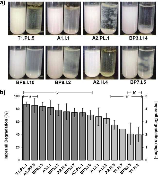
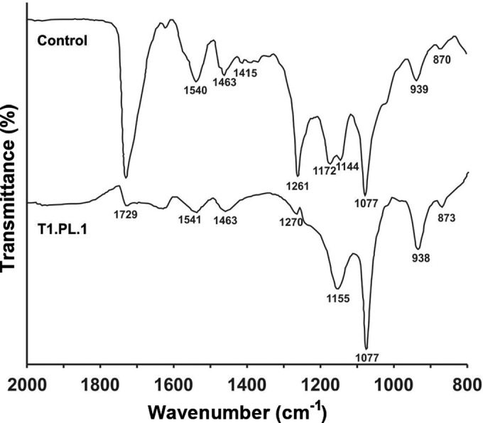
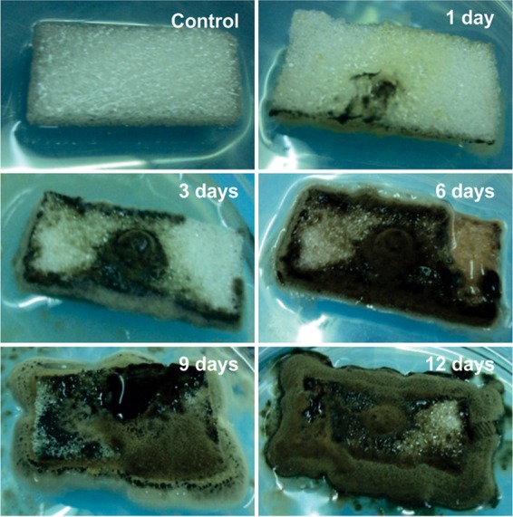
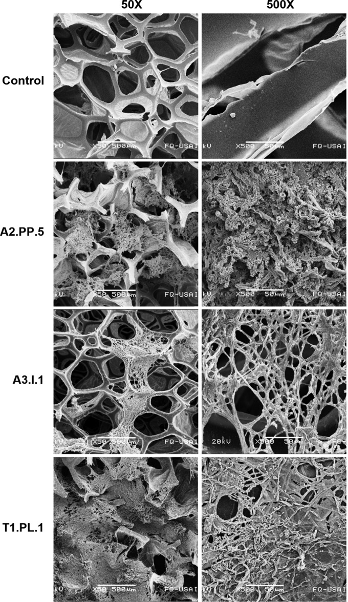
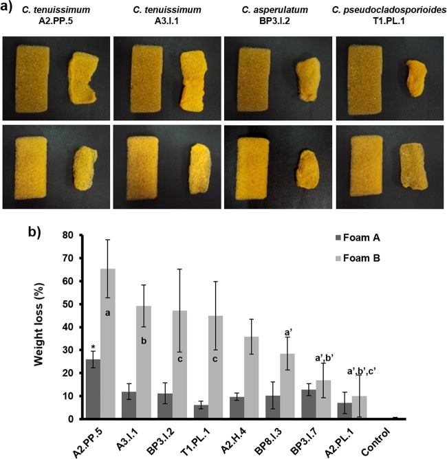
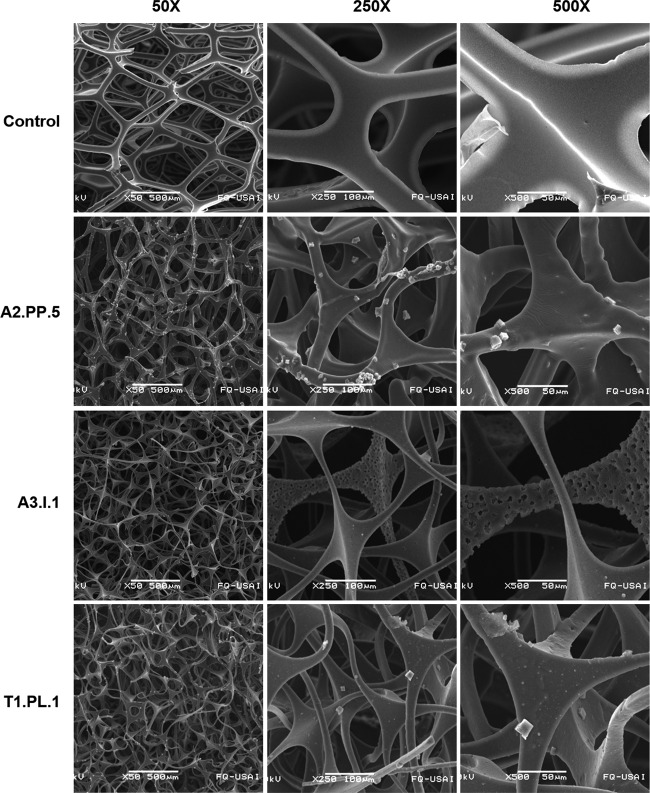
Polyurethane (PU) is widely used in many aspects of modern life because of its versatility and resistance. However, PU waste disposal generates large problems, since it is slowly degraded, there are limited recycling processes, and its destruction may generate toxic compounds. In this work, we isolated fungal strains able to grow in mineral medium with a polyester PU (PS-PU; Impranil DLN) or a polyether PU (PE-PU; Poly Lack) varnish as the only carbon source. Of the eight best Impranil-degrading strains, the six best degraders belonged to the Cladosporium cladosporioides complex, including the species C. pseudocladosporioides, C. tenuissimum, C. asperulatum, and C. montecillanum, and the two others were identified as Aspergillus fumigatus and Penicillium chrysogenum The best Impranil degrader, C. pseudocladosporioides strain T1.PL.1, degraded up to 87% after 14 days of incubation. Fourier transform infrared (FTIR) spectroscopy analysis of Impranil degradation by this strain showed a loss of carbonyl groups (1,729 cm(-1)) and N-H bonds (1,540 and 1,261 cm(-1)), and gas chromatography-mass spectrometry (GC-MS) analysis showed a decrease in ester compounds and increase in alcohols and hexane diisocyanate, indicating the hydrolysis of ester and urethane bonds. Extracellular esterase and low urease, but not protease activities were detected at 7 and 14 days of culture in Impranil. The best eight Impranil-degrading fungi were also able to degrade solid foams of the highly recalcitrant PE-PU type to different extents, with the highest levels generating up to 65% of dry-weight losses not previously reported. Scanning electron microscopy (SEM) analysis of fungus-treated foams showed melted and thinner cell wall structures than the non-fungus-treated ones, demonstrating fungal biodegradative action on PE-PU.
Importance: Polyurethane waste disposal has become a serious problem. In this work, fungal strains able to efficiently degrade different types of polyurethanes are reported, and their biodegradative activity was studied by different experimental approaches. Varnish biodegradation analyses showed that fungi were able to break down the polymer in some of their precursors, offering the possibility that they may be recovered and used for new polyurethane synthesis. Also, the levels of degradation of solid polyether polyurethane foams reported in this work have never been observed previously. Isolation of efficient polyurethane-degrading microorganisms and delving into the mechanisms they used to degrade the polymer provide the basis for the development of biotechnological processes for polyurethane biodegradation and recycling.
Copyright © 2016, American Society for Microbiology. All Rights Reserved.
Figures
References
-
- Plastics Europe. 2015. Plastics—the facts 2015. Plastics Europe, Frankfurt, Germany: http://www.plasticseurope.es/Document/plastics-the-facts-2015.aspx?FolID=2.
-
- Instituto Nacional de Estadística y Geografía. 2012. Anuario estadístico de los Estados Unidos Mexicanos 2011. Instituto Nacional de Estadística y Geografía, Aguascalientes, Mexico.
-
- Mahajan N, Gupta P. 2015. New insights into the microbial degradation of polyurethanes. RSC Adv 5:41839–41854. doi: 10.1039/C5RA04589D. - DOI
Publication types
MeSH terms
Substances
LinkOut - more resources
Full Text Sources
Other Literature Sources
Medical
Miscellaneous
