Review
. 2016 Dec 2:226:117-127.
doi: 10.1016/j.virusres.2016.05.031. Epub 2016 Jun 15.

Cellular entry of the porcine epidemic diarrhea virus

Affiliations
Review

Cellular entry of the porcine epidemic diarrhea virus

Wentao Li et al. Virus Res. .

Abstract

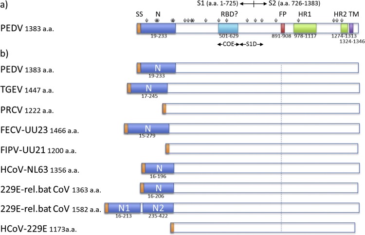
Porcine epidemic diarrhea virus (PEDV), a coronavirus discovered more than 40 years ago, regained notoriety recently by its devastating outbreaks in East Asia and the Americas, causing substantial economic losses to the swine husbandry. The virus replicates extensively and almost exclusively in the epithelial cells of the small intestine resulting in villus atrophy, malabsorption and severe diarrhea. Cellular entry of this enveloped virus is mediated by the large spike (S) glycoprotein, trimers of which mediate virus attachment to the target cell and subsequent membrane fusion. The S protein has a multidomain architecture and has been reported to bind to carbohydrate (sialic acid) and proteinaceous (aminopeptidase N) cell surface molecules. PEDV propagation in vitro requires the presence of trypsin(-like) proteases in the culture medium, which capacitates the fusion function of the S protein. Here we review the current data on PEDV entry into its host cell, including therein our new observations regarding the functional role of the sialic acid binding activity of the S protein in virus infection. Moreover, we summarize the recent progress on the proteolytic activation of PEDV S proteins, and discuss factors that may determine tissue tropism of PEDV in vivo.

Keywords: Coronavirus; Membrane fusion; PED; PEDV; Porcine epidemic diarrhea virus; Proteolytic activation; Receptor interaction; Sialic acid; Sialic acid binding; Spike; Virus; Virus entry; Virus tropism.


Figures

Fig. 1
Fig. 1
Schematic presentation of spike proteins of PEDV and related alphacoronaviruses. a) The PEDV S protein. The S1 and S2 subunits, the signal peptide (SP, residues 1–18), the N-terminal domain (N, residue 19–233), regions containing neutralizing epitopes (COE, residues 499–638 and S1D, residues 636–789), the fusion peptide (FP; residues 891–908), two heptad repeat regions (HR1, residues 978–1117 and HR2, residues 1274–1313), the transmembrane domain (TM, residues 1324–1346; predicted by TMHMM server) and predicted N-glycosylation sites (ψ; predicted by the NetNGlyc server) are indicated. b) N-terminal domains (N) in the spike proteins of PEDV and related alphacoronaviruses. Schematic representations of the spike proteins are shown of a number of viruses belonging to Alphacoronavirus genus including PEDV strain CV777 (GB: AAK38656.1), TGEV strain Purdue P115 (GB: ABG89325.1), PRCoV strain ISU-1 (GB: ABG89317.1), FECV strain UU23 (GB: ADC35472.1), FIPV strain UU21 (GB: ADL71466.1), HCoV-NL63 (GB: YP_003767.1), HCoV-229E related bat CoV with one N domain (GB: ALK28775.1), HCoV-229E related bat CoV with two N domains (GB: ALK28765.1) and HCoV-229E strain inf-1 (GB: NP_073551.1). Spike proteins are drawn to scale and aligned at the position of the conserved fusion peptide indicated by the dashed line. Signal sequence is indicated in orange.
Fig. 2
Fig. 2
Generation and characterization of a recombinant PEDV (PEDV-SΔN) with a large 215-residues long deletion in the N-terminal region of the S protein. a) Amino acid sequence alignment of the N-terminal region of the spike proteins of PEDV variants. The N-terminal region (residues 1–294) of the spike protein of the cell culture adapted DR13 strain (caDR13; GB: JQ023162.1) was aligned with the corresponding spike protein sequences of the caDR13 PEDV-SΔN recombinant described in this study and two earlier described PEDV variants with deletions in the N-domain − PC177 (GB: AKR05184.1) and Tottori2 (GB: BAR92898.1) − as well as of the PEDV-GDU strain (GB: KU985229) and PEDV-UU strain (GB: KU985229). Alphahelical (‘H’) and betasheet ('E') secondary structural elements were predicted using the JPRED4 server (http://www.compbio.dundee.ac.uk/jpred/). Conserved residues and gaps are indicated in the alignment using the ‘*’ and ‘-‘ symbols, respectively. Signal peptide and cysteine residues are marked in grey and black, respectively. b) Schematic representation of the recombinant PEDV genomes carrying the wildtype spike (SWT) gene (PEDV-SWT-GFP) or the SΔN gene (PEDV-SΔN-GFP). For both viruses, the S proteins carried a C-terminal Flag-tag peptide (VQDYKDDDDK) and the ORF3 gene was substituted by the GFP gene (Wicht et al., 2014b). Left panel displays pictures of Vero cells infected with PEDV-SWT-GFP or PEDV-SΔN-GFP taken by fluorescence microscopy. c) Western-blot analysis of the S protein of recombinant PEDV-SWT-GFP and PEDV-SΔN-GFP. Recombinant viruses propagated in Vero (CCL81) cells in the absence of trypsin were semi-purified by sedimentation through a 20% (w/w) sucrose cushion, subjected to Western blot analysis, after which S proteins were detected with an anti-Flag monoclonal antibody (Sigma). d) Multi-step growth kinetics of PEDV-SWT-GFP and PEDV-SΔN-GFP. Vero cells were inoculated with recombinant PEDV (MOI = 0.01) for 3 h in the absence of trypsin, after which the inoculum was replaced by fresh culture medium, following a previously described procedure (Wicht et al., 2014b). Virus infectivity in culture medium was determined at different times p.i. (4, 12, 24, 36, 48, 60, 72, 96 or 120 h p.i.) by a quantal assay on Vero cells from which TCID50 values were calculated.
Fig. 3
Fig. 3
PEDV virions of different strains vary in their hemagglutination activity. a) Virions of the PEDV-GDU strain but not of the PEDV-UU or PEDV-caDR13 strain are able to agglutinate human erythrocytes. The HA assay was performed according to a previously described procedure (Park et al., 2010) and the influenza A virus (A/California/07/2009) was taken along as a positive control. Two-fold serial dilutions of viruses (start dilution 1 × 107 TCID50/ml) were made in 50 μl phosphate buffered saline supplemented with 0.1% bovine serum albumin. Neuraminidase (from Arthrobacter ureafaciens, Sigma, cat.no. 10269611001) treated (10 mU ml−1 at 37 °C for 2 h) or mock treated human erythrocytes were washed with phosphate buffered saline. 50 μl erythrocyte suspension (0.5%) was mixed with 50 ul of each virus dilution in V-shaped 96 wells plates and incubated for 2 h on ice after which the wells were photographed. Virus dilutions are indicated above the plate. Wells positive for hemagglutination are encircled in red. b) The Fc-tagged S1 protein of the PEDV-GDU strain but not that of the PEDV-UU or PEDV-caDR13 strain is able to agglutinate human erythrocytes. To enhance the sensitivity of the S1-based hemagglutination assay, we premixed 5 μg of S1-Fc proteins with 1 ul of protein A-coupled, 200nm-sized nanoparticles (nano-screenMAG-Protein A beads; Chemicell GmbH, cat.no. 4503-1) to increase the avidity of S1-Fc proteins for sialic acids on the erythrocyte surface. The influenza A virus hemagglutinin glycoprotein ectodomain (A/California/07/2009, GB: ACP41953.1) fused to the human Fc portion (IAV-HA-Fc) was taken along as a positive control. The start dilution of IAV-HA-Fc and PEDV-S1-Fc was 5 μg and two-fold serial dilutions of virus-nanoparticle mixtures were tested as decribed above.
Fig. 4
Fig. 4
Dependency of PEDV on cell surface sialic acids for infection of Vero cells varies between strains. a) Vero cells were pretreated with PBS or PBS containing neuraminidase (100 mU ml−1; Sigma, cat.no. 10269611001) at 37 °C for 2 h. Cells were subsequently inoculated with PEDV-GDU, PEDV-UU or influenza A virus (IAV-WSN) (Baggen et al., 2016) at a multiplicity of infection of 5, 1 or 5, respectively, for 1 h at 37 °C in the presence of trypsin. Inoculum was removed and cells were washed thrice with fresh medium and further incubated at 37 °C in medium supplemented with 1% fetal calf serum. At 14 h p.i. cells were fixed and infected cells were visualized by immunofluorescence staining using a mouse monoclonal antibody detecting PEDV nucleocapsid protein (BioNote, Republic of Korea). b) Percentage of infected cells (relative to PBS-treated) was calculated by counting the infected cells in 10 x microscopic fields. Statistical significance was assessed by unpaired one-tailed Student’s test (* = P<0.01). The infection experiments were repeated three times, and representative images are shown.
Fig. 5
Fig. 5
Contrasting trypsin-dependency of recombinant PEDV carrying spike proteins from different PEDV isolates. a) PEDV requires proteolysis to activate membrane fusion. The pictures show a PEDV-infected cell fusing with neighbouring cells upon trypsin addition. Snapshots from a time-lapse movie of an infected cell culture taken at 3, 16, 27 and 45 min after addition of trypsin to the culture medium are shown in the following order: upper left, upper right, lower left and lower right, respectively. Pictures represent an overlay of fluorescence (virus encoded-mEGFP labelled cells [green] and Hoechst 33342 stained nuclei [blue]) and DIC images). b) Recombinant viruses encoding spike proteins of PEDV-CV777, PEDV-GDU and the cell-culture adapted DR13 isolate (GB: AF353511, AFP81695.1, JQ023162.1, respectively) were generated in an isogenic background by targeted RNA recombination, as described before (Wicht et al., 2014b). In all recombinant viruses the ORF3 gene was replaced by that of GFP. Vero cells were inoculated with recombinant viruses (MOI 0.1) in the absence or presence of soybean trypsin inhibitor (SBTI) or trypsin, or in the presence of both, as indicated. The cells were fixed at 15 h post infection, nuclei were stained with DAPI (blue) and nuclei and infected cells (GFP; green) were visualized by fluorescence microscopy.

Similar articles

Cited by

References

    1. Baggen J., Thibaut H.J., Staring J., Jae L.T., Liu Y., Guo H., Slager J.J., de Bruin J.W., van Vliet A.L., Blomen V.A., Overduin P., Sheng J., de Haan Xander C.A., de Vries E., Meijer A., Rossmann M.G., Brummelkamp T.R., van Kuppeveld F.J. Enterovirus D68 receptor requirements unveiled by haploid genetics. Proc. Natl. Acad. Sci. U. S. A. 2016;113:1399–1404. - PMC - PubMed
    1. Belouzard S., Chu V.C., Whittaker G.R. Activation of the SARS coronavirus spike protein via sequential proteolytic cleavage at two distinct sites. Proc. Natl. Acad. Sci. U. S. A. 2009;106:5871–5876. - PMC - PubMed
    1. Belouzard S., Millet J.K., Licitra B.N., Whittaker G.R. Mechanisms of coronavirus cell entry mediated by the viral spike protein. Viruses. 2012;4:1011–1033. - PMC - PubMed
    1. Burkard C., Verheije M.H., Wicht O., van Kasteren S.I., van Kuppeveld F.J., Haagmans B.L., Pelkmans L., Rottier P.J., Bosch B.J., de Haan C.A. Coronavirus cell entry occurs through the endo-/lysosomal pathway in a proteolysis-dependent manner. PLoS Pathog. 2014;10:e1004502. - PMC - PubMed
    1. Chang S., Bae J., Kang T., Kim J., Chung G., Lim C., Laude H., Yang M., Jang Y. Identification of the epitope region capable of inducing neutralizing antibodies against the porcine epidemic diarrhea virus. Mol. Cells. 2002;14:295–299. - PubMed

Publication types

MeSH terms

Substances